The Future of Epilepsy Treatments at the Comprehensive Epilepsy Center
From advanced diagnostics to advanced therapeutic and surgical treatments to cutting-edge clinical trials, the neurologists, neurosurgeons, neuropsychologists, nurse practitioners, nurses and other staff at the Comprehensive Epilepsy Centers at Children’s Wisconsin (Children’s) and Froedtert Hospital specialize in unique, personalized plans that can control seizures and improve quality of life.
“Approximately 3.5 million people are living with epilepsy in this country, and every one of those cases is different. That makes treating it extremely expensive for most healthcare systems,” says Manoj Raghavan, MD, PhD, director of the Comprehensive Epilepsy Program, medical director of the Magnetoencephalography Program and MCW professor of neurology and neurosurgery. “MCW, Froedtert and Children’s stand out because we offer the resources, equipment and providers that other systems in Wisconsin can’t.”
Working in conjunction with MCW faculty, both centers are certified Level 4 by the National Association of Epilepsy Centers (NAEC) – the highest level of certification possible – and offer the state’s only magnetoencephalography (MEG) program. MEG is a noninvasive tool that works like a three-dimensional electroencephalogram (EEG) to monitor brain function and track where seizures begin.
But that’s far from the only thing bringing the future of treatment to today’s patients.
Implanting the Future
For one patient who experienced frequent, drug-resistant seizures on both sides of her brain – making traditional surgery impossible – the team implanted a Responsive Neurostimulation (RNS) System in May 2022. The RNS device monitors brain activity, and when a possible seizure is detected, it immediately provides stimulation to the area of the brain where seizures arise in an attempt to disrupt the seizure.
The patient and her family saw immediate improvement, as her seizures dropped from approximately once per week to once per month or fewer, and her family hasn’t needed to use rescue medication (fast-acting drugs with anticonvulsant effects that are administered during seizures to stop or reduce the activity) since the implant.
“Nothing has increased quality of life like the RNS,” says LouAnn Counihan, the patient’s mother. “My daughter used to be tired all the time and we didn’t know why. Information from the RNS showed that she was having numerous seizures that we weren’t observing. Since the implant, her fatigue and moods are dramatically improved.”
Due to the combination of the patient’s improvements and the ability to know what’s happening during a seizure (via the information the RNS device provides), the family is planning to move to a new home that will offer their daughter the opportunity to live more independently and require less direct care.
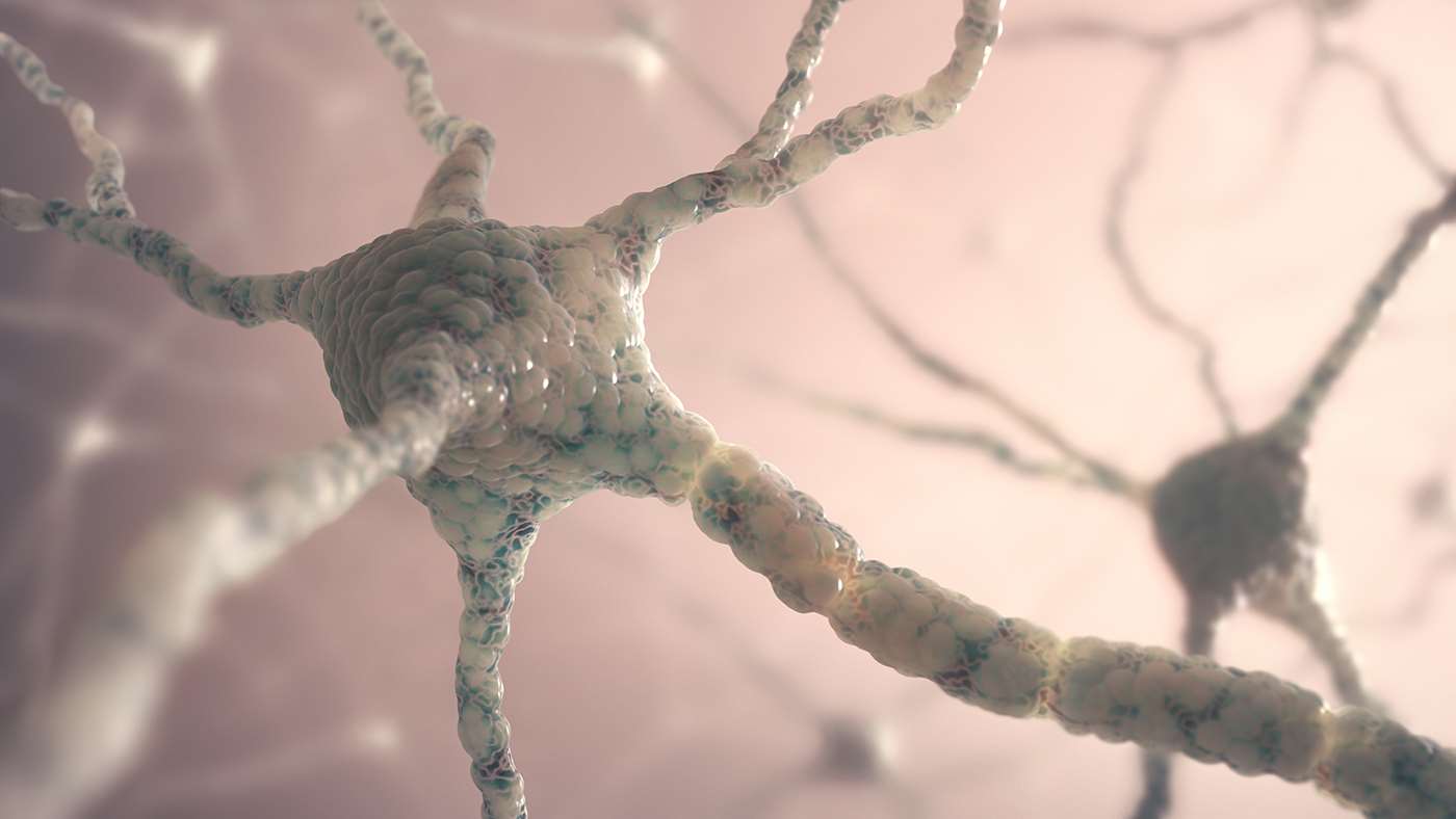
Meanwhile, additional research underway at MCW includes recruiting patients for a study on implantation of inhibitory cells into the hippocampus.
The majority of neurons in the brain are excitatory, meaning that they activate other neurons to further transmit electrical signals. Inhibitory cells do the opposite. Researchers hope to determine whether the inhibitory cells may be able to make neurons involved in a seizure less likely to fire, thus lessening the intensity and frequency of seizures and improving outcomes.
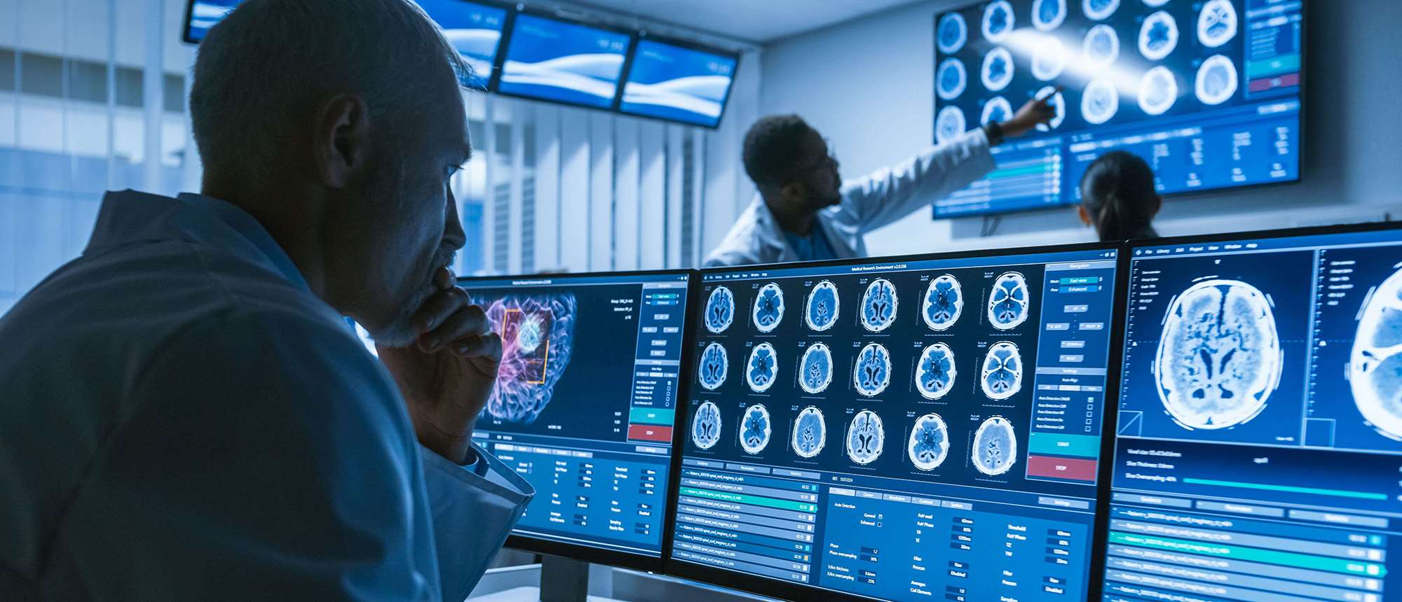
Next-Level Mapping of the Brain
Approximately two-thirds of people living with epilepsy can be treated with medication that will control their seizures. For the other third, however, surgery often is needed to improve their quality of life. For example, resecting (removing) the source of seizures can prevent seizures or limit their spread within the brain, thereby also limiting what patients experience or feel with their seizures. Before that can happen, however, vital areas of brain function must be perfectly mapped.
Research that was pioneered at MCW helped develop functional MRI (fMRI), which can help locate vital areas of the brain prior to surgery, as well as predict outcomes for language and memory capabilities.
Functional mapping with direct stimulation of the brain can be used during surgery to identify key brain functions in real time. For example, if seizure activity occurs near a portion of the brain vital to necessary functions – speech, skin sensations, ability to read, etc. – fMRI can help identify and protect those areas.
Other tools critical to detecting seizure location include positron emission tomography (PET)/MRI, which combines information on the metabolic activity of the brain (using PET) with structural images of the brain (from MRI) to help identify potential causes or foci for seizures which may be subtle and difficult to identify.
Neuropsychological testing provides a detailed neurocognitive picture of brain functions such as memory. This testing also may provide information regarding areas of the brain from which seizures arise and define risks of potential surgical procedures.
After reviewing all of these data to optimize outcomes, the epilepsy team can develop an individualized treatment plan designed to preserve function and control seizures. The neurosurgical team makes use of tools, including robotic-assisted surgery, laser ablation and stereoelectroencephalography (SEEG) to better understand and ultimately treat seizures.
Building for Tomorrow
“Seizures during critical times of a child’s development can reduce cognition later in life,” says Dr. Sean Lew, Mardak/Vandenberg Family Chair of Pediatric Neurosurgery at Children’s, MCW professor of neurosurgery, and director of epilepsy surgery at Froedtert Hospital and Children’s. “Thus, it’s important that we’re focused on reducing or eliminating seizures as much, and as early, as possible to maximize long-term quality of life.”
Every Tuesday, MCW faculty from the adult and pediatric epilepsy teams meet to review the latest cases and discuss the best options for treatment.
For each case, they consider the long-term effects of each treatment option and what tomorrow may hold for the person living with epilepsy, as well as for their respective caregivers.
The result is the future, today. And tomorrow.
Following the “Jennifer Aniston Neuron”
In 2005, neuroscientist Rodrigo Quian Quiroga, PhD – when looking for the areas of the brain that cause epileptic seizures – discovered what he called the “Jennifer Aniston Neuron,” which lighted up each time his research subject saw an image of the actress Jennifer Aniston or heard her name. This discovery of a neuron that is linked to a particular concept was a major milestone in understanding how the mind works.
Using a new intracranial research program, scientists at MCW have found that similar processes occur for many different brain stimuli. This work may even help uncover how one’s brain creates a memory.
Researchers in the lab of Hernan Rey, PhD, MCW assistant professor of neurosurgery, working with MCW neurosurgeon and epilepsy specialist Sean Lew, MD, at the Epilepsy Monitoring Unit at Froedtert Hospital, are implanting a series of specialized electrodes in deep areas of the brains of patients with refractory epilepsy.
During intracranial tests, specialized electrodes track brain activity as close as possible to an individual neuron. A computer immediately compiles the data and produces results that allow Dr. Rey to interpret brain processes.
As their seizures cannot be controlled by medication, these patients require invasive recordings to identify the area of the brain where their seizures originate. While undergoing these recordings, Dr. Rey and his team give patients numerous behavioral tasks to perform.
As neurons in their brains respond to each task, the activity is tracked via microwires that extend beyond the end of a standard clinical probe. A computer compiles the activity, response time, intensity and more, and processes the data almost immediately.
The data then allows Dr. Rey to interpret a variety of brain processes, including memory, attention and decision-making by reviewing how different areas of the brain react during the experimental tasks.
“We already know that certain neurons in the hippocampus are part of memory creation,” shares Dr. Rey. “This research puts us on the leading edge of finding how the hippocampus and other parts of the brain respond so we can better understand how these neural networks connect. We’re studying the memories that make us, us.”
Dr. Rey hopes this work ultimately may identify epilepsy biomarkers that could lead to clinical trials of more efficient treatments, improved margins for resection and better indicators for treatment options, as well as reduced side effects.
Check Out Our WINS!
The Wisconsin Institute of Neuroscience (WINS) – initially created in 2022 as the MCW Neuroscience Institute through a partnership among MCW, Froedtert Hospital, Children’s Wisconsin and the Clement J. Zablocki Veterans’ Administration Medical Center – was launched in January 2023 to assert MCW’s national presence in neuroscientific research and treatment.
 WINS strives to enhance basic and translational neuroscience through multidisciplinary, world-class clinical care, unique clinical trials, groundbreaking research and innovative education.
WINS is led by Shekar Kurpad, MD, PhD, Sanford J. Larson Professor and chair of neurosurgery at MCW.
The WINS name reflects the integration of the extraordinary capabilities, resources and clinical expertise of its four partners into a singular set of offerings.
The name also highlights the regional and national authority WINS brings to the neurosciences
– Chris Combs
Recommended


