
Keck Functional Imaging Laboratory (KFIL)

 The X-ray imaging system was designed for both dynamics planar X-ray angiography and micro-CT. This provides our group with the flexibility to acquire data on mass transport phenomena as well as detailed volumetric morphology. In both modes, emphasis was placed on high spatial resolution, on the order of tens of microns. Specifically, the system can acquire high frame rate planar images and short scan time volumetric data, which require high temporal resolution and high data throughput in addition to relatively low noise, large dynamic range, and a precision x-ray source and detector. The instrumentation also allows for precise manipulation of the specimen and detector position relative to the X-ray source providing fine controlled magnification and accurate selected regional imaging for data collection in real time.
The KFIL has additional functional imaging capability to study metabolic processes or other physiologic systems. Portable Gamma cameras are utilized for complimentary studies and can be combined with pin-hole collimation to provide high-resolution emission images. The same specimen can be analyzed with both X-ray and Gamma-ray modalities without repositioning. This provides added capabilities to record and merge functional and anatomic data.
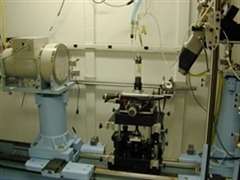 Animal studies are facilitated by various life support and physiologic apparatus housed in the imaging suite. Ancillary equipment is in close proximity to the specimen stage and has been designed to allow for unrestrained articulation of the animal, organ, or tissue specimen.
To date the KFIL has been utilized for a number of disease related studies including, pulmonary hypertension, vascular remodeling, bronchial angiogenesis, ventilator induced injury, instilled surfactant transport, renal function and stone formation, tumor perfusion, gastro-esophageal reflux disorder, coronary collateral vessel growth, vascular stent implantation, lymphatic transport, cranial suture ossification, bone strength and development. The microfocal X-ray scanner that enables the examination of blood vessels and other anatomy as small as several microns is the only scanner of its kind and the only microCT system in the Milwaukee area. The KFIL is a unique facility dedicated to basic biomedical research combining imaging modalities that enables morphometric and functional analysis of a range of basic physiological investigations and provides insight into the structural and metabolic mechanisms involved in health and disease.
The KFIL’s research portfolio continues to grow and the laboratory welcomes additional collaboration studies. If you are interested in utilizing the imaging lab for current or future projects, please contact Ann Clough, PhD, at ann.clough@marquette.edu for more information.
Example images and renderings from Keck Functional Imaging Laboratory


