Ophthalmology & Visual Sciences at the Eye Institute

Case Study 5 - CC: Girl rubbing her R eye after trauma

all
Patient Visit

Patient History
HPI:

The patient is a 2 year-old Caucasian girl who presents to the ED after falling from bed at 4:30 am and landing on a glass nightstand which shattered on impact. Mom checked her and found no bruises or bleeding. The next morning mom noticed her rubbing the R eye several times. She also keeps that eye closed by squinting all the time. She will not allow mom to look at the eye.

Past Ocular History:
No hx of ocular surgery, trauma, amblyopia, strabismus.

Ocular Medications:
None

Past Medical History:
Healthy child. Born at term.

Surgical History:
None

Past Family Ocular History:
Negative for blinding diseases.

Social History:
Lives with mother. No smoking in the house.

Medications:
None

Allergies:
None

ROS:
No recent illnesses.

Ocular Exam

Visual Acuity (cc):
OD: Open eye to command but keeps eye closed most of the time. Central, unmaintained.
OS: Central, steady and maintained

IOP (tonoapplantation):
OD: Not performed
OS: Not performed

Pupils:
Irregular, peaked OD; Round OS. No APD.

Extraocular Movements:
Full OU. No nystagmus.

Confrontational Visual Fields:
Not performed.

External:
Normal-appearing orbital structures, both sides.

Slit Lamp:

Lids and Lashes Normal OU
Conjunctiva/Sclera Trace diffuse hyperemia OD; normal OS
Cornea 3 mm corneal laceration at 8 o’clock in horizontal configuration, extending from the limbus towards the visual axis OD; clear OS
Anterior Chamber Flat OD; deep and quiet OS
Iris Peaked, iris to wound OD; normal OS
Lens Normal OU
Anterior Vitreous Normal OU, brief view due to patient cooperation
Dilated Fundus Examination:
OD No good view.
OS Clear view, no VH; CDR 0.1 with sharp optic disc margins; flat macula with normal foveal light reflex; normal vessels and peripheral retina
Other:
None
Diagnosis and Discussion

Diagnosis
Globe rupture R eye

Discussion

Differential Diagnosis:
This patient presents with a full-thickness cornea laceration in the R eye after trauma. Other etiologies for full-thickness corneal defects include wound dehiscence (if patient has a history of prior corneal surgery) or corneal perforation due to dellen, scleritis, neurotrophic keratopathy, corneal ulcer, chronic topical anesthetic abuse or environmental exposure (crack smoking). Suspect child abuse in any child with unexplained globe rupture and initiate the appropriate evaluation.

Definition:
A full-thickness laceration is a through-and-through defect of the eye wall (corneal or scleral) that causes the globe to be open to the environment. Other instances of open globe include blunt trauma resulting in rupture of previous surgical wounds and perforating trauma (with an entry and exit wound of the eye). It is important to inquire as to the nature of trauma or foreign body (vegetable matter vs. glass vs. metal).

Examination:
Typical symptoms of globe rupture are pain, redness, tearing and decreased vision. Exam findings include: other lid or orbital trauma (ex. lid laceration – examination of the eye under a lacerated eyelid is imperative), conjunctival hemorrhage and/or swelling, corneal abrasion and/or laceration, flat or shallow anterior chamber, anterior chamber cell, hyphema, peaked pupil and/or iris rupture, high or low IOP, lens opacity, vitreous hemorrhage, retinal tear and/or detachment, choroidal rupture and/or intraocular foreign body. The eye exam should be comprehensive, but limit the manipulation of the affected eye (ex. avoid IOP check). Ensure the unaffected eye is examined (with dilation). The seidel test is a method of detecting aqueous humor leakage from the cornea by applying fluorescein to the site of laceration in the cornea. If positive a yellow fluid will be seen under cobalt blue light. An orbital CT scan is indicated in most cases to identify any foreign body, to examine the sclera for shape abnormalities (posterior ruptures can be missed on clinical exam) and to examine the orbital bones for fractures. In instances when the posterior pole can't be examined a B-scan could indicate intraocular foreign body, retinal detachments or vitreous hemorrhage but care should be placed not to put undo pressure in the eye.

Treatment:
Prompt surgical repair of the laceration is necessary to restore ocular integrity. The patient should receive anti-emetic, pain and antibiotic intravenous medication, a shield over the affected eye and tetanus prophylaxis while waiting for surgical repair. Outcomes are unpredictable and depend on the extension of the injury. An important prognosticator of final visual acuity is presenting visual acuity. Injured eyes require lifelong monitoring. Sympathetic ophthalmia is an inflammatory condition where the immune system attacks the uninjured eye, most likely due to systemic exposure of ocular antigens. In eyes with no possibility of vision recovery, enucleation can decrease the risk of immune activation and also resolves severe eye pain. Lifelong monocular precautions - using protective eye wear at all times - is recommended.

Self-Assessment Questions
  1. What elements of the ocular examination would suggest globe rupture?
  2. What elements of the ocular examination should be avoided when suspecting ocular rupture to prevent dehiscence of ocular content?
  3. What is the 1st step that should be taken once a globe rupture is diagnosed?


References/Resources:
Ehlers JP, Shah CP, Fenton GL, et al. The Wills Eye Manual, 5th edition. Lippincott Williams, & Wilkins: Philadelphia, PA. 2008.

Friedman NJ, Kaiser PK, Pineda R. The Massachusetts Eye and Ear Infirmary Illustrated Manual of Ophthalmology, 3rd edition. Saunders Elsevier: China. 2009.

Kanski JJ. Clinical Ophthalmology: A Systematic Approach, 5th edition. Butterworth Heinemann: St. Louis, MO. 2003.

University of Iowa Department of Ophthalmology and Visual Sciences

Self-Assessment Answers

1. What elements of the ocular examination would suggest globe rupture?

f. All of the above

2. What elements of the ocular examination should be avoided when suspecting ocular rupture to prevent dehiscence of ocular content?

a. Removing an embedded foreign body in the cornea

Any foreign body embedded in the cornea or other part of the eye should be removed in the operating room at the time of globe rupture repair.

3. What is the 1st step that should be taken once a globe rupture is diagnosed?

b. Place a shield over the affected eye

It is imperative to protect the affected eye to prevent expulsion of intraocular content due to patient rubbing the eye. The other answers should be performed soon after.

Contact Ophthalmology

For patient care inquires, call us at (414) 955-2020 or use MyChart. Email is for research and education inquiries only.

Eye Institute Location

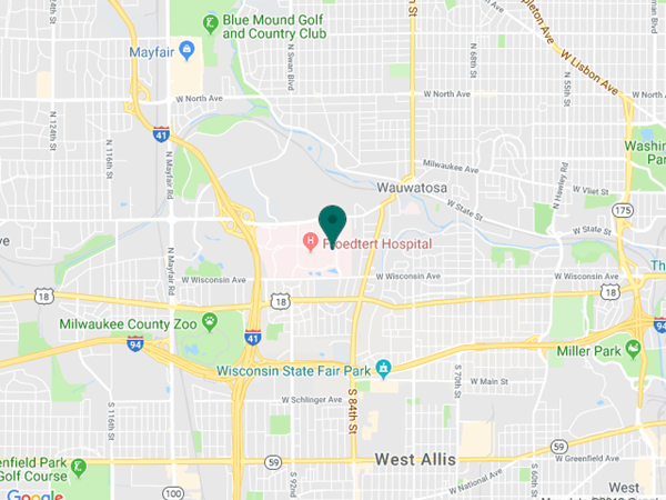
925 N. 87th St.

Milwaukee, WI 53226


Appointments

(414) 955-2020

(414) 955-6166 (fax)


Continuing Medical Education

Taylor Schmidt

taschmidt@mcw.edu

(414) 955-2088


Medical Education Coordinator

Ophth-Residency@mcw.edu


Associate Director of Development - Ophthalmology

Sarah Walker

sarawalker@mcw.edu

Refer to Us - Consultation requests

Patient Referral Form (PDF)

Fax to (414) 955-0136


Emergent Requests

Within 48 hours call

(414) 955-2020


Research

Vesper Williams

vewilliams@mcw.edu

(414) 955-7862


Advanced Ocular Imaging Program

aoip@mcw.edu

(414) 955-2647


Eye Institute Executive Director (Administrator)

Shannon Dreier
sdreier@mcw.edu

Eye Institute Google map location