
Engineering Better Clinical Imaging Diagnostics
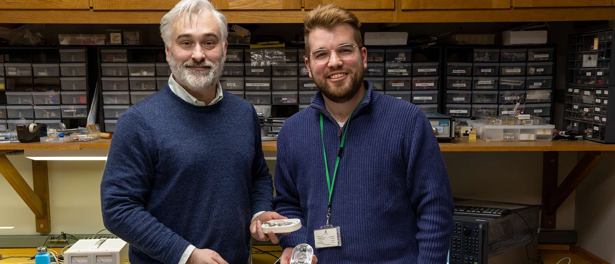
Ask Jason W. Sidabras, PhD, about his first life plan and he’d mention his passion for building the electronics in guitar pedals.
Originally, Dr. Sidabras set out to pursue that goal, studying electrical engineering technology at the Milwaukee School of Engineering. While there, he was pulled in a different direction: antenna design.
Today, as an assistant professor in the Department of Biophysics at the Medical College of Wisconsin (MCW), Dr. Sidabras designs and builds the antennas in MRI (magnetic resonance imaging) machines.
MRIs are used to create images of soft tissues in a patient’s body. To do so, a person lies in an MRI machine which contains a strong magnet that aligns hydrogen atoms within the water and fat in the body. Next, a brief pulse of radio waves disrupts this alignment. As the atoms relax back to their aligned state, they produce a signal that the scanner detects. Using additional magnetic fields to determine where those signals came from, a computer reconstructs the detailed image of internal tissues.
Amid this complex chain of physics, Dr. Sidabras engineers the radiofrequency antennas – the component that emits the radio waves and captures the resulting signal. When the antennas capture a weak signal, radiologists and physicians have a hard time interpreting the data – in other words, understanding what they are seeing inside a patient.
“The joke is that the spectroscopists need to look at the wiggles and my job is to make the wiggles bigger,” he says.
Dr. Sidabras believed the key breakthrough would come from redesigning the antenna itself, which traditionally takes the form of a metal, coiled loop. He was right. Around a decade ago, he discovered that when he paired two particular coil geometries together, they significantly outperformed a standard loop.
Engineering a Better Patient Outcome
After spending years optimizing the design, Dr. Sidabras and his then graduate student and now postdoctoral researcher Kyle Johnson, PhD ‘24, refined the technology into a practical tool that can be used on commercially available MRI scanners. With the ability to boost MRI signal by at least 2.5 times, their work has the promise of being a breakthrough with strong clinical potential.
That means for patients the benefits are two-fold:
- First, a larger signal means a cleaner, clearer image of internal tissues.
- Second, and as a direct result, a better image allows clinicians to spot a biological anomaly – or the absence of one – more quickly, which translate into less time in the MRI machine.
This can be particularly useful for children, whose patience can wane when asked to remain still.
“Don’t get me wrong, I like the physics, but watching the design evolve into a tool that can really change someone’s life, their prognosis, or their diagnosis, that’s something that really excites me about the technology,” says Dr. Johnson, adding that he and Dr. Sidabras hope to commercialize the technology and bring it directly to patients.
From Magnetic Resonance to Radiation Detection
Beyond MRIs, Dr. Sidabras applies his technology to investigate protein structure, dynamics, and function. In fact, much of his published research focuses on advancing these methods.
In a more unconventional application and backed by a $1.5 million dollar grant, Dr. Sidabras explores how the technology can help in the event of a nuclear disaster. When nuclear radiation hits the body, it breaks sulfur bonds, which generate a detectable signal. The device measures this signal and determines how much radiation the body absorbed.
Sulfur is found in keratin, a protein in nails. Researchers hope to incorporate the technology into a small, portable device that analyzes a person’s nails to detect radiation exposure on the spot. Current methods require a blood draw sent to a lab for analysis.
“The idea is that if anything catastrophic happens, we need a way to quickly assess people’s radiation dose and tell them they will be fine if they stay home or that they need to go to the hospital,” says Dr. Sidabras.
Mentorship and Music
A freedom-to-explore mentorship style characterizes Dr. Sidabras's lab. To him, making messes is part of the scientific process.
“One of the things that I try to teach is to fail fast and fail often because that’s when you build your intuition,” he says.
Dr. Sidabras's first love, music, still hums in the background. A part-time musician who performs in three rock bands, he can often be found quietly jamming in the lab, bringing a creative spark to his scientific work.
“A love of music permeates a lot of conversations in the lab because it’s not always about science, it's about connecting with one another,” says Dr. Johnson. “If you develop that connection, the work and professional relationship becomes stronger.”
That spirit of curiosity and collaboration has helped drive an innovative technology now reaching beyond his lab.
“I think the idea that what I’m doing does not sit in these four walls anymore is quite invigorating,” says Dr. Sidabras.
Recommended


