

Medical College of Wisconsin Researchers Utilize Retinal Phantom to Standardize Cutting-Edge Eye Imaging in International Phase III Clinical Trial
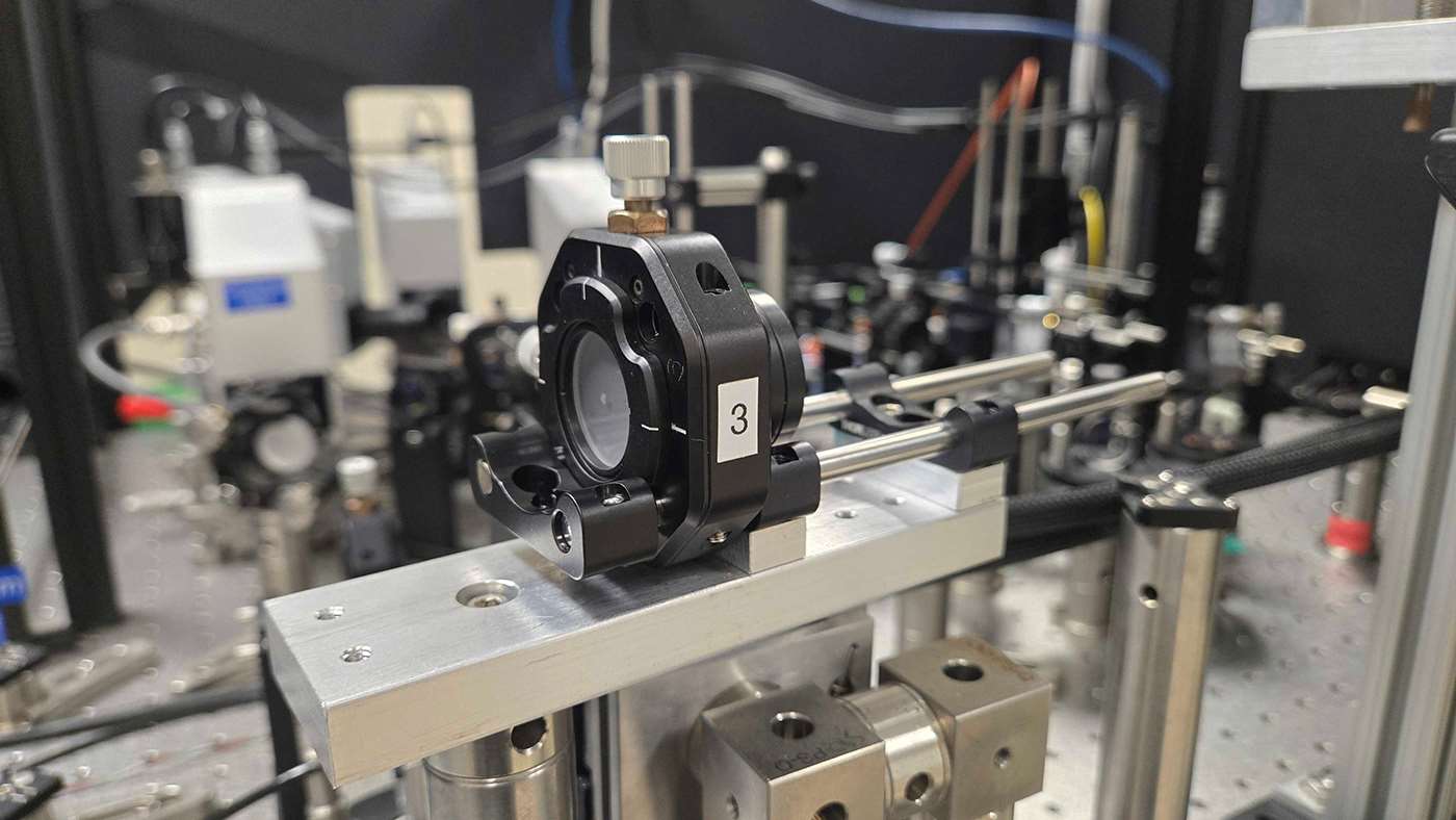
When a patient with retinitis pigmentosa is imaged using Adaptive Optics Scanning Light Ophthalmoscopy (AOSLO) in London and another is imaged in Milwaukee, how do researchers know that the data they see is consistent across highly customized imaging equipment?
That is the question vision scientists at the Medical College of Wisconsin (MCW) aimed to answer in the newly released paper, “Use of a Retinal Phantom to Facilitate Adaptive Optics Scanning Light Ophthalmoscopy Imaging in a Multicenter Clinical Trial,” published in the August issue of Translational Vision Science & Technology, an ARVO journal. This work was performed through the AOSLO Resource Center grant – co-directed by Dr. Joseph Carroll at MCW and Dr. Jacque Duncan at the University of California, San Francisco – as part of the larger NIH-funded NAC Attack clinical trial (NCT05537220).
“We have imaging systems with very high-resolution capabilities, but currently, they're all independently constructed, and a lot of them have different designs,” explains Joseph Kreis, the paper's leading author and graduate student in the Dennis P. Han, MD Advanced Ocular Imagining Program (AOIP) at MCW. “We wanted to leverage high-resolution retinal imaging capability across multiple sites for this clinical trial, but we didn't have a way to compare data across unique systems.”
Standardizing current AOSLO systems to a single design wasn’t a realistic option. Many of these devices have been custom-built by individual labs over years, and renovating them would mean adding cost, delay, and technical burden to research teams who often run these trials with little dedicated funding. Instead, the team utilizes a retinal phantom, that any existing system could image the same way, without requiring labs to change their equipment or workflow.
The retinal phantom, which uses a 3D printed design mimicking cone photoreceptors at the cellular level, was developed by Anant Agrawal, PhD, Research Electrical Engineer and Daniel X. Hammer, PhD, Acting Director of Division of Biomedical Physics, at the FDA’s Center for Devices and Radiological Health (CDRH), in collaboration with University of Maryland professor Ryan D. Sochol, PhD, Director of the Bioinspired Advanced Manufacturing (BAM) Laboratory, and former student Ian Rosenthal. Six identical retinal phantoms, all printed on the same 3D printer, were sent out to the six AOSLO imaging sites for the NAC Attack phase III multicenter clinical trial, ensuring every site evaluated its imaging system against the exact same standard. The paper presents phantom imaging data collected over nearly two years, starting before enrolling any patients. During the trial, the phantom was imaged each time a study site sees a patient for AOSLO imaging, or once every six months if there was a gap in visits. Data collected at least twice a year allowed researchers to monitor systems and flag any significant changes. The NAC Attack clinical trial itself is designed to determine if the oral medication N-Acetylcysteine will help slow the progression of vision loss in patients with retinitis pigmentosa.
NAC Attack is the first clinical trial using adaptive optics imaging at multiple independent study sites, and deploying retinal phantoms to each site gives researchers the confidence to say that patient imaging results are the same regardless of imaging site. For patients, that could mean shorter clinical trial periods, faster FDA approval of new drugs, and the ability to image more patients across the globe. For researchers, the phantom allows them to compare data collected from multiple custom systems in a consistent manner, ultimately making clinical trials more efficient.
“The phantom has also revealed trends in the AOSLO systems that site coordinators were not aware of, which has led to conversations about improving lab operations and what to watch for when imaging patients in a clinical trial,” says Kreis. “Day-to-day research allows a lot of flexibility, but a trial running four-plus years demands consistency. You want to know you're not negatively impacting the data, especially when seeing the same patient repeatedly over several years.”
Not only does the retinal phantom help standardize imaging done with existing AOSLO systems but go-to-market AOSLO systems have emerged that could eventually become standard imaging devices in clinics, like optical coherence tomography (OCT) is today. Retinal phantoms help maintain consistent device performance – a path the paper’s authors hope to make viable in clinical settings.
“The bigger picture is that the ability to detect changes at a single-cell level means outcomes can be measured in a much shorter time than a typical clinical trial requires,” Kreis shares. “That leads to shorter trials, which means studies can enroll more patients, test drugs on a shorter timescale, and get therapies approved and accessible faster, increasing throughput for the number of drugs, sites, and patients that can be tested. Not having to wait three years for someone to lose 10 letters of visual acuity, when we can see meaningful change in two years, or even two months, lets us get people the treatment they need sooner.”