
MCW Researcher Studies the Hidden Life of DNA's Packaging Using NMR
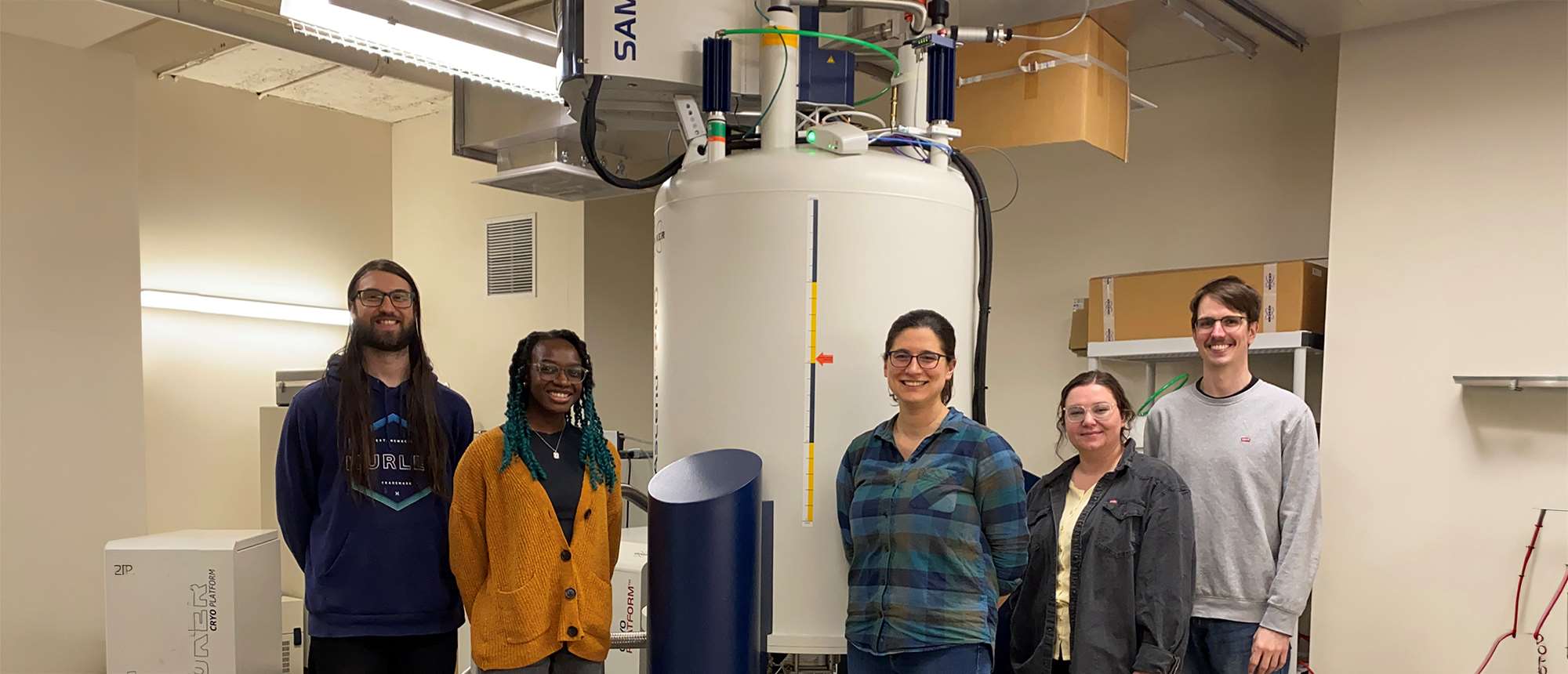
Emma Morrison, PhD, associate professor of biochemistry at the Medical College of Wisconsin (MCW), loved to visit her father’s chemistry lab when she was a child. He would nick some coffee filters from the break room and give Dr. Morrison and her sister pipettes to fill with the liquid fluorescence that he used for cancer diagnostics. The two girls would dribble designs onto the filters, then he’d grab his portable black light so they could see their creations glow in the dark.
“That was the most fun part of going there,” she says.
Dr. Morrison's father was her role model, so she decided to major in chemistry as an undergraduate at Johns Hopkins University. Soon, though, she found herself excited by biophysics, a field that combines chemistry, physics, and biology.
“It's all very overlapping and interdisciplinary,” she says. “You can call yourself a physical biochemist, a chemical biologist, or a biophysicist. I consider myself a molecular biophysicist.“
The molecules that she studies are proteins, and she uses a technique called Nuclear Magnetic Resonance (NMR) spectroscopy to understand their structure and motion.
NMR uses a powerful superconducting magnet to align the atomic nuclei in a molecule, then it employs a sequence of radiofrequency pulses to nudge those aligned nuclei. How they respond to different sequences of pulses reveals information about the molecule's structure and dynamics.
“NMR is unique among structural biology approaches in that you can look at biomolecules moving in solution, similar to a cellular environment,” Dr. Morrison says. “Then you can study their dynamics – their motion – across different time scales from picoseconds (one trillionth of a second) to seconds.”
Shape-shifting Proteins
Dr. Morrison attended Washington University in St. Louis for her PhD. She worked in the lab of Katherine (Katie) Henzler-Wildman, PhD, who had a project using NMR to study EmrE, a protein in the bacteria E. coli. EmrE is a multidrug resistance transporter, meaning it can bind to a small-molecule drug like an antibiotic then pump that drug out of the cell so the bacteria survive.
What made EmrE interesting was its ability to change between two shapes as it moved a broad range of drug molecules from one side of the cell membrane to the other, aiding in antibiotic resistance.
“I was really intrigued by the question of how one protein can not only bind to such a wide range of (drugs),” she says, “but also have this global conformational change when it's bound to each of them.”
She measured the speed of the shape switching and the strength of the connection as it bound to different drugs. She found the switching rate varied enormously depending on the drug, with the weakest-bound drug actually causing the fastest switching.
“It was a really cool project,” says Dr. Morrison. “Katie had a lot of enthusiasm and it was contagious, so I decided to stick with NMR. I don't regret it.”
Understanding the Role of ‘Reader’ Proteins
Next, Dr. Morrison began a postdoc at the University of Iowa with Catherine Musselman, PhD, who was studying histones – proteins that form spool-like structures called nucleosomes. A cell's DNA, which would stretch six feet if uncoiled, wraps around these spools.
Proteins called “writers” can deposit chemical tags on histones, and then other proteins called either “readers” or "erasers" interact with those tags, either binding or removing them.
Using NMR, Dr. Morrison started out looking at those "reader" proteins. In doing so, she realized that nucleosomes themselves were doing more interesting things than just sitting there waiting to be read.
For example, histones have tails that extend out from the nucleosome core. These tails could interact with nearby DNA by both folding back and attaching to the DNA in their own nucleosome or by grabbing DNA from a nearby one.
In doing so, the histone tails affect gene expression. For instance, when a tail grips some DNA, it helps block access to it, silencing genes. But when writer proteins add chemical tags to the tail, the grip can loosen, allowing the DNA to open and genes to be turned on.
“That's the idea that I brought with me to MCW when I started my lab,” she says. “That the nucleosome and its components have an active role in the so-called histone language rather than just being a platform that other proteins interact with.”
Histone Tails Drive Organization
Dr. Morrison’s lab is focused on how histone tails contribute to something called chromatin, which is the name for the entire package of nucleosomes in a cell, which number in the tens of millions.
One area of chromatin organization that she’s interested in is called phase separation. It’s based on the idea that cell nuclei may be organized into distinct zones – not by physical membranes, as with other cellular compartments – but by regions of chromatin that spontaneously separate from each other.
Dr. Morrison thinks the histone tails are driving this process – because each nucleosome has ten tails, capable of grabbing onto DNA from multiple neighboring nucleosomes simultaneously.
This capability means they form a sprawling network that naturally clumps into phase-separated regions. Because chemical tags can loosen or tighten the tails' grip on DNA, those modifications likely determine how these regions form and dissolve.
In one paper, she found the specific pattern of charged amino acids in a histone tail determines what kind of phase-separated zone forms, and that tweaking that code by adding modifications changes the zone's properties.
Aspiring to Help Cancer Patients Through Chromatin Function
Dr. Morrison’s work could help make new drugs that target a cell’s nucleus more effectively. Researchers are targeting the nucleus, especially the proteins that read, write, and erase chemical tags on chromatin, because many diseases, especially cancer, involve genes being incorrectly turned on or off.
But if those researchers think of chromatin itself as just a passive backdrop, they’re potentially missing a whole layer of biology that impacts disease development, and their drug may not work as intended.
“I think it's important to understand these fundamental processes in order to truly understand how to rationally design new therapeutics within the cell’s nucleus,” says Dr. Morrison.
Thus, Dr. Morrison’s work could ultimately benefit cancer patients, which gives it a connection to her father’s work that she finds satisfying.
She says he worked on DNA probes that target chromatin, improving cancer diagnostics, and making a real difference in patients’ lives.
“We ended up in a similar research space,” she says, “but I can only aspire to influence people as much as I think he has.”
For him, the most important influence may have been on her. “My parents,” she said with a smile, “are very proud.”
Recommended


