MCW Researchers Team Up to Decode How a Common Virus Can Harm the Developing Brain
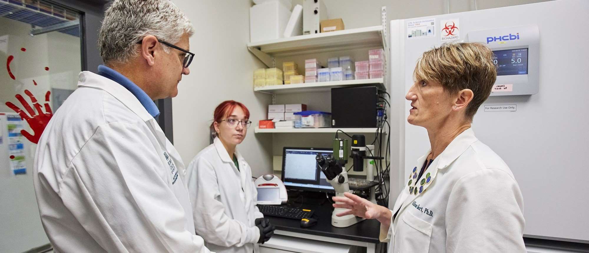
She grows mini brains known as organoids to better understand neurological diseases while he teases apart how viruses rewire cells. Together – Allison Ebert, PhD, professor of cell biology, neurobiology and anatomy and director of the Neuroscience Research Center, and Scott Terhune, PhD, professor of microbiology & immunology – are researching cytomegalovirus, a common virus that can cause serious birth defects and is especially devastating when it affects brain development.
“There’s a lack of diagnostic tests and no approved therapeutics for uncovering and treating the infection in utero," says Dr. Terhune. “It’s very, very complex.”
Drs. Terhune and Ebert are digging into how cytomegalovirus upends the structure and function of developing neurons and neuron-protecting glial cells in the brain. Their 2023 ground-breaking study in the peer-reviewed journal, mBio showed that the virus doesn’t just target immature brain cells, as previously believed, but also more mature cells that have developed into functioning neurons.
Now with a $1 million gift from the Stead Foundation to study infection and brain health, and a $3.4 million National Institutes of Health grant, they will spend the next five years moving beyond what happens in single brain cells to include the surrounding tissue as well.
That’s important, Dr. Terhune says, because when a virus infects a cell, the infected cell changes neighboring cells as well.
“This new grant is allowing us to really take that next step in the level of complexity to understand how the infection is impacting a tissue's development,” he says.
Replicating Tissue for Ethical Research
 Dr. Ebert will create that tissue by growing neural organoids. To make them, she takes human skin or blood cells and turns them into what are known as “induced pluripotent stem cells.”
Like embryonic stem cells, these cells can grow into almost any cell type in the body – but they’re created without the ethical concerns tied to using embryos. Dr. Ebert turns these simple cells into more complex brain cells such as neurons and glial cells to form a mini-nervous system in a Petri dish.
“There's no other way to truly model human brain development other than utilizing these types of cells,” she says. “You can literally watch them grow in a dish and then test the perturbations that are specific to human disease at a very early developmental stage.”
In this case, the "perturbation" is infection by cytomegalovirus. That’s where Dr. Terhune’s expertise comes in. He is fascinated by viruses, pointing out that while they are simple in structure – just a handful of components – they have a big effect on host cell architecture.
 “They're not complex,” he says, “but boy, when they enter a complex system, they change everything.”
How they do that is linked to the proteins they make and which parts of the host cell they bind to. The cytomegalovirus makes more than 200 proteins and the brain cells they invade make tens of thousands. To unravel those connections, Dr. Terhune uses a technique called peptide mass spectrometry.
“You infect cells with the virus, pull out the viral protein of interest, find out what it's associating with, and then you make a hypothesis about what the protein is likely doing,” he says. “From there, you can try blocking the interaction to see if it has an effect on disease development.”
Tracing Distant Effects of the Virus
Dr. Terhune’s collaboration with Dr. Ebert means he can study the virus’s protein connections in the exact type of brain cells that it invades.
“It’s made the research much more impactful,” he says.
The researchers joined forces 12 years ago when one of Dr. Terhune's graduate students, Tarin Bigley, MD, PhD ‘16, now at Washington University School of Medicine, learned Dr. Ebert was new to campus and had the ability to grow brain cells.
“So much of this research has been student-driven,” says Dr. Ebert. “Shortly after I got here, Tarin came asking, ‘Oh, can we infect some neural progenitor cells with cytomegalovirus?’ I said ‘Sure, I'm not a virologist, I have no idea what you're talking about, but let's give it a go.’”
With this latest round of funding, the two will employ spatial transcriptomics to illuminate the location of downstream effects of the viral protein interactions.
“We know from some of our data that even a distant cell that doesn't have active viral replication is still not normal,” Dr. Ebert says. “So, we're trying to figure out what the virus is actually doing that's causing that distant damage.”
Unravelling those complex interactions could then lead to both diagnostic tests and therapies for developing fetuses infected with the cytomegalovirus.
“To me,” says Dr. Terhune, “that would be a home run.”
Recommended


