
Medical College of Wisconsin Biochemistry Research Facilities & Shared Equipment
The Department of Biochemistry has several pieces of shared equipment and shared research facilities that are available for use.
BIAcore Instrumentation

About the BIAcore S200 SPR Instrument
BIAcore S200 SPR
The Biacore S200 SPR instrument can measure interactions of various sample types, from low molecular weight drug candidates to high molecular weight proteins (also DNA, RNA, polysaccharides, lipids, cells, and viruses) in various sample environments (e.g., DMSO-containing buffers, plasma, and serum).
Applications include:
- Fragment screening and LMW drug discovery
- Kinetic and affinity determination
- Competition assays
- Epitope mapping of antibodies
- Thermodynamics
The high sensitivity and low baseline noise of the Biacore S200 facilitate reliable affinity, kinetic, and fragment-screening data. The Biacore S200 is optimized for small molecule library screening, with automated software to evaluate non-specific binding, identification and prioritization of binders, and calculation of association (ka) and dissociation (kd) rate constants and affinity (KD). The Binding Level Screen function provides a rapid overview of small molecule libraries, automatically identifying fragments above a defined cut-off level. The predefined template for Binding Level Screen was developed explicitly for a 384-well plate format, allowing 384 compounds to be screened in less than 16 hours.
The S200 software also offers a range of tools for kinetic analysis, including analysis of single-cycle kinetics where several concentrations of an analyte are injected in the same cycle. Multicycle injections can still be analyzed, but by eliminating surface regeneration between injections, single-cycle kinetics simplifies the analysis of targets that are unstable or difficult to regenerate. Single-cycle kinetics also reduce analysis time. By utilizing the predefined templates for kinetics, thirty different analytes can be run in as little as 16 hours.
The Biacore S200 also simplifies competition studies to validate small molecules' interaction site with the capability to use ABA injections. The ABA injection mode allows two different solutions to be injected over the surface in the same cycle consuming much less inhibitor than traditional SPR experiments. In addition, the ABA injection feature can help identify ternary complex formation with multiple ligands.
The Biacore S200 instrument is housed in the Biochemistry Department. It is available to all Medical College of Wisconsin faculty and staff who are trained and can demonstrate proficiency on the instrument. Training and consultation are available on an appointment basis.
For more information on SPR, please see the following links provided by the manufacturer, Cytiva:
- Main Biacore page: Get started with SPR-interaction analysis
- Free on-line course: Get started with Biacore SPR assay
BIAcore 3000
The Biacore 3000 SPR instrument is no longer operational. The computer with the control and evaluation software, and data will be maintained for users.
Usage Fees:
(Note: A training period of no less than 4 hours is required before you can work unassisted on the instrument. BIAcore chips and special reagents are not included in the fee.)
Training
Academic Users: $50/hour | Industrial/Non-Academic Users: $75/hour
Unassisted Use
Academic Users: $12.50/hour | Industrial/Non-Academic Users: $40/hour
Consultation (experimental design, data evaluation)
Academic Users: $50/hour | Industrial/Non-Academic Users: $75/hour
For more information contact:
Adriano Marchese, PhD
(414) 955-4191 | amarchese@mcw.edu
Ya Zhuo, PhD
(414) 955-4192 | yazhuo@mcw.edu
NMR Instrumentation
About Biomolecular NMR at MCW
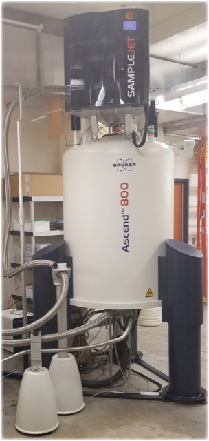
The NMR Facility is an interdepartmental research service unit located in the Biochemistry Department. High-field NMR spectroscopy is a powerful technique for the study of biomolecular structure and dynamics. The facility provides service for routine 1D and 2D NMR, and also provides consultation and collaborative assistance with the acquisition and analysis of multidimensional, multinuclear protein NMR spectra. The facility maintains four Bruker NMR spectrometers (Avance III 500 MHz, two Avance-III HD 600 MHz, and Avance-Neo 800 MHz) which are operated and maintained by Dr. Francis Peterson (Professor of Biochemistry and NMR facility manager) in a dedicated 2500 ft2 facility in the Health Research Center basement. Each NMR instrument is equipped with a 1H/15N/13C cryoprobe for ultra-high sensitivity in biomolecular applications. The 500 MHz instrument also has 19F detection, and the 800 MHz cryoprobe is equipped with three-axis XYZ pulsed-field gradients. SampleJet robots for automated NMR acquisition of samples in 96-well sample racks are installed on the 600 and 800 MHz instruments. One 600 MHz instrument is configured with a 1.7mm 1H/15N/13C cryoprobe (~60 µL sample size) and dedicated to chemical fragment screening and other automated multi-sample/high-throughput NMR applications. For long-term projects, the facility provides training for instrument operation and data analysis to investigators and research personnel. The facility operates on a fee-for-service basis and is open to faculty of the Medical College of Wisconsin and outside researchers.
Rates:
- $9/hour for trained users using the 500 or 600 MHz instruments
- $12/hour for trained users using the 800 MHz instrument
- $25/hour for assisted use
- Inquire for industrial rates
For more information contact
Brian Volkman, PhD (414) 955-8400, bvolkman@mcw.edu
Francis Peterson, PhD (414) 955-5777, fpeterso@mcw.edu
Paul Kerber, MS (414) 955-7363, pkerber@mcw.edu
Macromolecular X-Ray Crystallography Facility


Biochemistry Shared Research Instrumentation
The Biochemistry Department maintains several instruments for isolation and physical characterization of biomolecules and detection of their interactions. All are located on the second floor of the TBRC and include:
Jasco J-710 Circular dichroism spectropolarimeter
The Jasco J-710 circular dichroism (CD) spectropolarimeter is equipped with a thermally regulated sample compartment. Monitoring of the far-UV and/or near-UV CD spectra can provide valuable information about the secondary structure, thermal stability, or conformational state of a protein.
Contact: Blake Hill, rbhill@mcw.edu
MicroCal VP - Isothermal Titration Calorimetry
The MicroCal VP-ITC is capable of measuring heat evolution as little as 0.4 nanoJ/sec. This instrument is suitable for the studies of protein-ligand and protein-protein interactions and provides the biochemists with reliable measurements of binding constants in the range of 103-109 M-1 as well as the enthalpy and stoichiometry of interactions. ITC is a preferred technique to demonstrate the interaction between newly discovered binding partners in vitro.
Contact: Brian Smith, brismith@mcw.edu