GettyImages-650160957-hero

Medical College of Wisconsin Stroke, Neurointervention, and Critical Care

Interventional neurology is a subspecialty field of medicine that combines highly specialized skills from the disciplines of neurology, neurosurgery, and neuroradiology to offer minimally invasive therapies for disorders of the brain and spine. The Froedtert & the Medical College of Wisconsin team in Milwaukee serves patients with brain aneurysms, arteriovenous malformations (AVM), dural arteriovenous fistulas (dAVF), carotid stenosis, stroke, and other disorders of the blood vessels of the brain and spine. Modern stroke care requires that physicians with special expertise make rapid determinations of a patient's status and of the optimal management. Treatment options vary from routine admission for observation and work-up to immediate intracranial thrombolysis. Our Division is fortunate to have several fellowship trained Neurologists specializing in stroke and both neurointensive and neurointerventional care, who can provide state-of-the-art treatments. The clinical expertise assembled in the Department of Neurology had resulted in JCAHO certification of Froedtert Hospital as the first Primary Stroke Center in Wisconsin in 2004.

Meet Our Stroke Neurointervention & Critical Care Team

headshot

Hatim Attar, MD

Assistant Professor

Specialty: Neuro Hospitalist

headshot

Jeffrey R. Binder, MD

Professor

Specialty: Stroke

headshot

Diane S. Book, MD

Associate Professor

Specialty: Stroke/Memory

headshot

Marek Cierny, MD

Assistant Professor

Specialty: Stroke

headshot

Ann K. Helms, MD, MS

Professor

Specialty: Stroke/Hyperbaric

headshot

Marc A. Lazzaro, MD, FAHA

Associate Professor

Specialty: Neuro Intervention/Stroke

headshot

Caitlin Moore, MD

Assistant Professor

headshot

Gregory E. Rozansky, MD

Assistant Professor

Specialty: Neuro Critical Care

Advanced Practice Providers

Department Info

all
Our Team

The Division of Neurointervention at Froedtert Hospital and the Medical College of Wisconsin is home to four neurointerventionalists with training in Neurology, Neurocritical Care, Vascular Neurology, and Endovascular Surgical Neuroradiology. The group provides consultation, evaluation, and follow-up for patients with a broad range of neurovascular conditions including those amenable to minimally invasive image-guided and catheter-based therapies. Located in Wisconsin’s largest academic health center, the Milwaukee Regional Medical Center, the Division of Neurointervention has become a leading referral center for neurovascular care in the Midwest. Extensive participation in landmark national clinical trials has positioned the Division of Neurointervention at the forefront of emerging therapies that are advancing the field. This group of physicians focuses largely on acute care in an inpatient sitting

  • Hatim Attar, MD
  • Jeffrey Binder, MD
  • Diane Book, MD
  • Mark Cierny, MD
  • Pramod Gupta, MD
  • Ann Helms, MD
  • Vijay Johnson, MD
  • Marc A. Lazzaro, MD
  • Gregory Rozansky, MD
  • Christopher Southwood, MD

Advanced Practice Providers

  • Nichole Brenton, NP
  • Melissa Carroll, NP
  • Cayla Jansen, NP
  • Stacie Foy, NP
  • Erin Grueneberg, DNP
  • Emily Klar, PA-C
  • Pooja Lemke, NP
  • Preska Patel, NP 
  • Beth Schultz, NP

Neurointervention Clinical Fellows 2019-20

  • John Cox, MD
  • Christopher Southwood, MD

Vascular Clinical Fellows 2019-20

  • Gabriel Bonnell, MD

Neurocritical Care Fellows 2019-20

  • Amanda Willcox, MD
  • Gregory Rozansky, MD

Fellowship Program Coordinator

Jenny Her | (414) 955-0643

Referrals

Our office can be reached by telephone Monday through Friday from 8 a.m. to 4:30 p.m. For referrals call (414) 805-6053. For emergency physician-to-physician transfers, please call (414) 805-4700.

Clinic appointments are held within the Froedtert West Clinics in the Neurosciences Research Center which is located on the 2nd floor of the Froedtert West Clinic building.

Locations & Directions

New patients are asked to bring the following:

  • Pertinent medical records
  • Prior imaging studies including CT, MRI, angiogram, or ultrasound studies.

Records may also be sent in advance to:

Nicole Turk or Judie Elliott
Department of Neurology
9200 W. Wisconsin Ave.
Milwaukee, WI 53226

Procedures are performed on the second floor of Froedtert Hospital in the Neurointerventional Angiography Suite using one of three biplane angiography machines.

Fellowships

The Division of Stroke, Neurointervention and Critical Care offers the following fellowships:

Clinical Trials
Clinical research is essential to advancing healthcare by defining efficacy in treatments and discovering safer and more cost-effective methods of delivering care to patients. The members of the Division of Neurointervention at Froedtert and the Medical College of Wisconsin share this vision and strive to be leaders in clinical trials. The Division of Neurointervention has participated in design and enrollment in numerous national clinical trials evaluating neuroendovascular therapies and devices and has been a top enrollment center in several multi-center randomized trials.

Current enrolling trials include:

The THERAPY Trial: The Randomized, Concurrent Controlled Trial to Assess the Penumbra System’s Safety and Effectiveness in the Treatment of Acute Stroke: Keywords: Stroke, Intervention

Penumbra Separator 3D US 510k Trial: Keywords: Stroke, Intervention

ACE: An Aneurysm Coiling Efficiency Study of the Penumbra Coil 400 System: Keywords: Aneurysm, Coiling, Intervention

DIAS-4: A randomized, double-blind, parallel-group placebo-controlled phase III study to evaluate the efficacy and safety of desmoteplase in subjects with acute ischemic stroke: Keywords: Stroke, Intervention

More information about ongoing neuroscience clinical trials can be found on the Froedtert Hospital website.
Stroke Program
Stroke programs provide comprehensive care to patients. From the emergency room to physical rehabilitation, stroke programs combine the skills of many different disciplines offering prevention, diagnosis, and treatment of stroke, and the rehabilitation of the patient.

Learn more

Selected Disorders and Conditions

all
Acute Ischemic Stroke

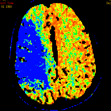
Acute stroke affects nearly 800,000 Americans annually making it the leading cause of disability and the 4th leading cause of death in the United States. About 80% of these strokes are due to a blockage of a blood vessel known as an “ischemic stroke.” This leads to inadequate blood flow to a part of the brain and causes a focal neurological deficit such as speech difficulty, numbness or weakness of an arm or leg, or vision loss. Timely restoration of blood flow to the brain is critical to prevent permanent damage to the affected brain tissue.

Time is brain, which is why rapid decisions are needed in the treatment of acute ischemic stroke. Patients experiencing an acute stroke require expedited diagnostic imaging of the brain and swift evaluation and management. Those who present within 4.5 hours from symptom onset and meeting specific criteria are eligible for a “clot busting” medication known as tissue plasminogen activator (tPA) administered through an IV. Patients who are not eligible for the IV medication may benefit from endovascular therapy using local administration of a medication at the clot in the blood vessel or clot removal with a device.

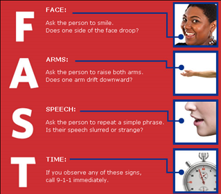
Knowing the signs and symptoms of stroke is important. A way to remember the stroke signs is F.A.S.T.:

  • Face: Ask the person to smile to determine if one side of the face droops.
  • Arms: Ask the person to raise both arms to determine if one drifts down.
  • Speech: Ask the person to repeat a simple phrase to determine if speech is clear or not.
  • Time: Call 911 immediately with any of these signs.

Act F.A.S.T. and call 911 immediately.

Acute Ischemic Stroke

Acute Ischemic Stroke

Arteriovenous Malformation

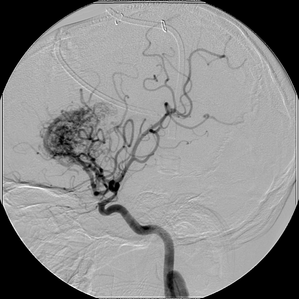
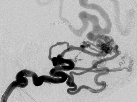
An arteriovenous malformation (AVM) is an abnormality of the blood vessels that may be present in the brain or spine and is believed to develop at the time of embryonic development. The malformation results in shunting of blood from arteries to veins, bypassing capillary blood vessels that serve normal brain tissue. The condition is rare and is thought to be present in about 0.1% to 0.2% of the population. The blood vessel abnormalities may not cause any significant neurologic problems and be discovered incidentally, or they may cause a wide variety of symptoms depending on the location. An AVM may be identified after headaches, a seizure, or rupture that causes bleeding. The workup for a patient with a suspected AVM often involves imaging studies such as CT or MRI of the brain, as well as catheter cerebral angiography to comprehensively define the abnormality. Various management strategies are then considered including observation, endovascular embolization, surgical resection, and gamma knife radiation.

Arteriovenous Malformation (AVM)Arteriovenous Malformation (AVM)

Arteriovenous Malformation (AVM)

Brain Aneurysms

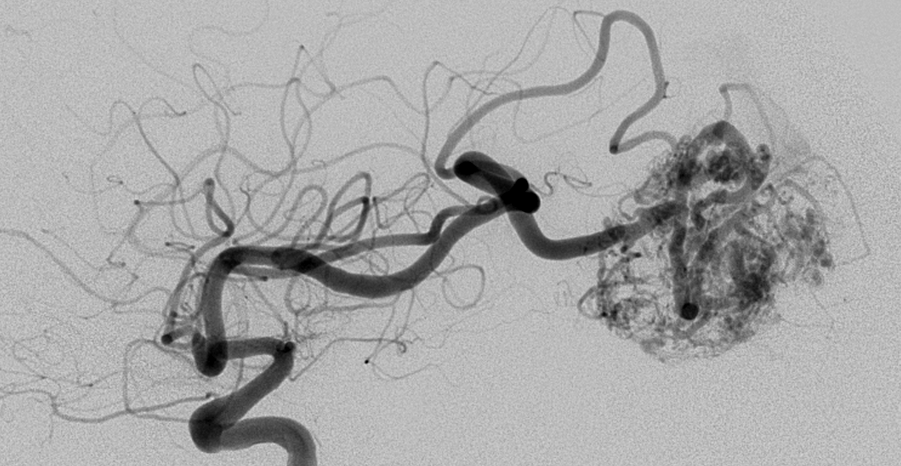
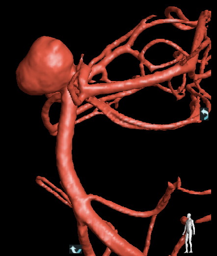
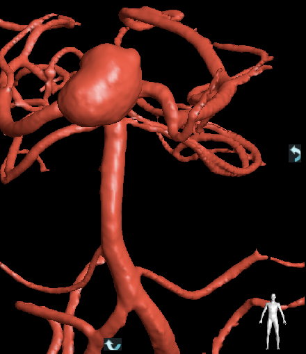
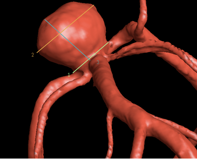
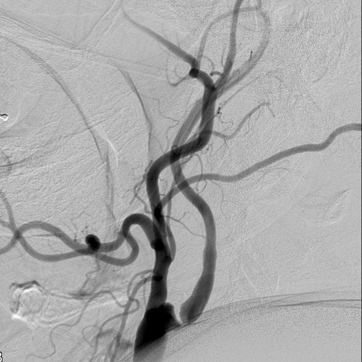
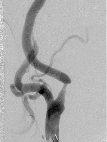
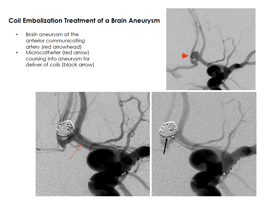
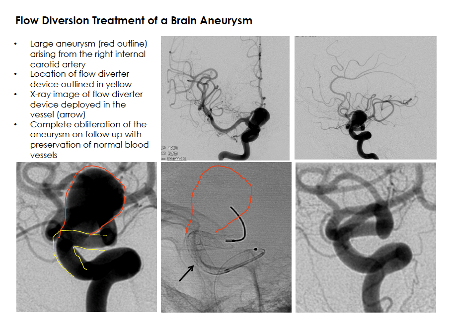
A brain aneurysm (cerebral aneurysm) is a bulge arising from the side of an artery wall in the head. It may be present from birth or develop after injury to the blood vessel. It has been estimated that up to about 5% of the population has a cerebral aneurysm. Symptoms of an aneurysm can include headaches or focal neurological deficits (such as a cranial nerve injury leading to difficulty with eye movement). Although rupture of an aneurysm is uncommon, the bleeding into the space surrounding the brain known as subarachnoid hemorrhage causes a severe headache and carries a high risk of severe neurologic injury and death. Risk factors for cerebral aneurysm growth include a family history, smoking, high blood pressure, and certain medical problems including polycystic kidney disease. Unruptured aneurysm treatment varies depending on several factors including the aneurysm shape, location, and a patient’s overall health. Management strategies include observation, endovascular coil embolization or blood flow diversion, and microsurgical clipping. A ruptured cerebral aneurysm is an emergency that requires urgent treatment.

Brain Aneurysm

Brain Aneurysm

Brain Aneurysm

Carotid Artery Stenosis

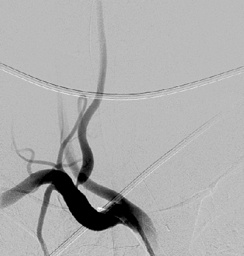
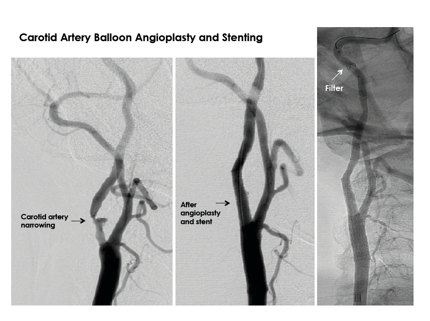
The carotid arteries are two large blood vessels in the neck that supply oxygenated blood to the brain. Carotid artery disease includes atherosclerosis which leads narrowing of the artery due to buildup of fatty substances and cholesterol deposits known as plaque. Stenosis, or narrowing, of these arteries can lead to stroke, a leading cause of death and disability in the United States. Signs and symptoms of carotid artery disease include transient ischemic attacks (TIA) or stroke. Patients may report changes in vision, strength, sensation, speech or other abnormalities. Evaluation of the carotid arteries can be performed with several non-invasive imaging methods including ultrasound, MRI, and CT. Management depends on various factors including the degree of narrowing, symptoms, and overall health. Treatment can include a combination of medicines to combat progression of plaque formation, lifestyle modifications, open surgery, or minimally invasive endovascular treatment.

Carotid Artery Stenosis

Carotid Artery Stenosis

Dural Arteriovenous Fistula
A dural arteriovenous fistula (dAVF) is an abnormality of the blood vessels that leads to shunting of blood from arteries to veins at an area of the tough covering over the brain (dura). The abnormality is very rare and is often associated with a prior traumatic injury. Patients may present with a variety of symptoms depending on the location, or the abnormality may be found incidentally when imaging is performed for an unrelated condition. Workup includes brain and spine imaging such as CT or MRI and often requires diagnostic catheter cerebral angiography for definitive diagnosis. Management options include observation, endovascular embolization, and microsurgical resection. Multiple factors must be considered to choose the optimal management strategy.

Dural Arteriovenous Fistula (dAVF)
Vertebral Artery Stenosis

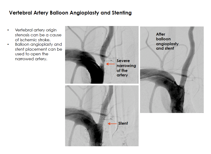
The vertebral arteries are a set of paired arteries that supply oxygenated blood to the back part of the brain. Atherosclerosis, or hardening of the arteries due to fatty substance deposition and plaque buildup, can lead to narrowing of the blood vessels. Patients may present with stroke or transient ischemic attack (TIA) symptoms such as a focal deficit of vision loss, dizziness, speech difficulty, or numbness or weakness of an arm or leg. Evaluation of the vertebral arteries can be performed with non-invasive imaging such as an MRI or CT scan. Patients with this condition can benefit from lifestyle changes and a medication regimen that will limit the progression of the narrowing. For patients who experience further narrowing and symptoms despite medications, some may benefit from an endovascular procedure to open the blocked artery.

Vertebral Artery Stenosis

Selected Procedures

all
AVM/AVF Embolization

Embolization of brain and spine arteriovenous malformations (AVMs) and arteriovenous fistulas (AVFs)

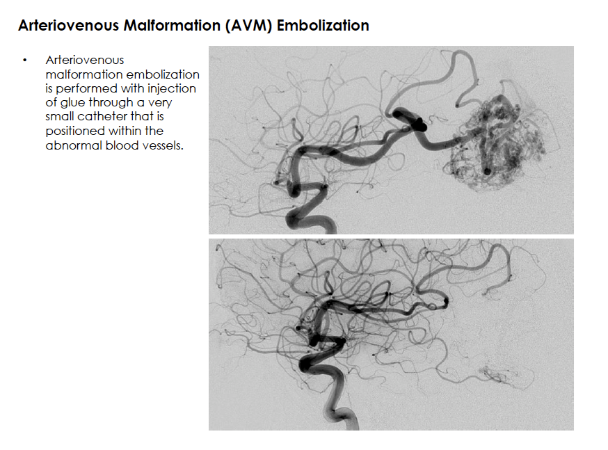
An arteriovenous malformation or dural arteriovenous fistula is an abnormality of the blood vessels that causes shunting of blood from an artery to a vein, bypassing normal tissue. This high-flow system may pose a risk for bleeding. Many characteristics of the abnormality may be important in deciding the best management approach. Endovascular embolization therapy is a method of injecting a “liquid embolic” or glue-like material into the target vessel to stop blood flow through the abnormality. A microcatheter (a very small hollow plastic tube) is carefully navigated to the abnormal connection between the artery and the vein. A glue material is then injected through the catheter to fill the abnormal collection of blood vessels, effectively closing the communication. The catheter is removed and the solidified glue remains within the blood vessels to permanently close the abnormality. This procedure may be performed alone or in conjunction with other treatments including open surgical resection or radiosurgery therapy.

Arteriovenous Malformation (AVM) Embolization

Brain Aneurysm Coiling and Flow Diverter Implantation

Some brain aneurysms can be closely observed with periodic surveillance imaging. For aneurysms that require treatment, endovascular therapy can be performed from inside the blood vessel. Access to the inside of the aneurysm is achieved with the same techniques used in diagnostic catheter cerebral angiography, by advancing a catheter (a hollow plastic tube) through an artery in the leg and navigating it to the blood vessels in the brain. After the catheter is positioned at the site of the brain aneurysm, several techniques are used to close the aneurysm and prevent any further blood flow entry. Coil embolization, or “coiling,” is the most commonly practiced endovascular treatment for brain aneurysms. In this procedure, a very small catheter is advanced into the aneurysm and platinum coils are sequentially introduced to create a tightly packed coil mass within the aneurysm, thereby promoting clot formation within the aneurysm, and preventing entry of blood flow. In some cases, a stent or balloon is required to remodel the neck of the aneurysm for safe coil placement and to promote healing. A balloon is temporarily inflated at the aneurysm neck, while a stent is permanently implanted to provide continuous support at the opening of the aneurysm. Newer technology and techniques now include flow diversion devices for endovascular aneurysm treatment. In this method, a mesh stent-like device is placed within the vessel and across the neck of the aneurysm to divert blood flow away from the aneurysm and down the normal course of the blood vessel. This promotes aneurysm closure and scaffolding for healing of the blood vessel wall. The rapid pace of technology development and evolution of techniques for endovascular repair of brain aneurysms will likely continue to advance safety and efficacy of minimally invasive therapies.

Coil Embolization Treatment of a Brain Aneurysm

Flow Diversion Treatment of a Brain Aneurysm

Carotid Artery Balloon Angioplasty and Stenting
Carotid artery narrowing is a known cause of ischemic stroke. Treatment options for selected patients include a minimally invasive endovascular approach to restore normal blood flow. In balloon angioplasty, a small balloon is temporarily inflated across the region of narrowing to expand the artery to normal diameter and restore blood flow. This procedure is usually accompanied by placement of a permanent stent over the narrowed region. During the procedure a temporary filter is placed in the internal carotid artery, downstream from the plaque, to capture debris that might be dislodged during the procedure.

Carotid Artery Balloon Angioplasty and Stenting
Carotid Cavernous Fistula Embolization
A carotid cavernous fistula can develop after trauma or spontaneously. Symptoms can include swelling and injury to nerves that control eye function. Imaging tests such as CT or MRI can provide clues to the diagnosis, but often a diagnostic catheter cerebral angiogram is necessary to confirm the diagnosis. Treatment options include endovascular therapy in which a small catheter is advanced into the blood vessels where the abnormal communication exists. Glue or a similar liquid embolic material is injected through the catheter and forms a plug at the abnormality to close the fistula.

Carotid Cavernous Fistula Embolization
Cerebral and Spinal Angiography
A diagnostic catheter cerebral or spinal angiogram is a procedure that is performed to acquire high-resolution images of the blood vessels of the head and neck or spine. This is also the foundation to all endovascular interventional procedures. After administering local anesthesia (usually lidocaine), an incision approximately 1/8 of an inch is made in the crease of the groin and a small sheath (a hollow plastic tube) is advanced into the femoral artery in the leg. A smaller catheter (a hollow plastic tube) is advanced through the sheath and navigated under x-ray guidance through the arterial system to the arteries that serve the head and the neck. Contrast dye is injected through the catheter into selected vessels and a series of x-ray images are rapidly acquired using two cameras positioned around the patient’s head. Catheter angiography affords the unique capability of evaluating dynamic and functional anatomy of the brain and spine with far greater resolution than non-invasive imaging such as CT or MRI. Characteristics of the circulation system including blood vessel anatomy and blood flow can be interpreted from the images.

The procedure is commonly performed with the patient awake but very sleepy, under “conscious sedation.” Sedation medications are administered to achieve patient comfort. During contrast injections, a patient may experience a sensation of warmth, flashes of lights, intermittent dizziness, or unusual tastes. These symptoms are well-tolerated, brief, and pass within seconds.

At the completion of the procedure, the catheter and sheath are removed, and pressure is applied to the leg or a small plug is placed to close the artery access site. The patient then recovers with flat bed rest for approximately 3 to 6 hours before returning home the same day.

Diagnostic Catheter Cerebral Angiography
Emergent Acute Stroke Therapy

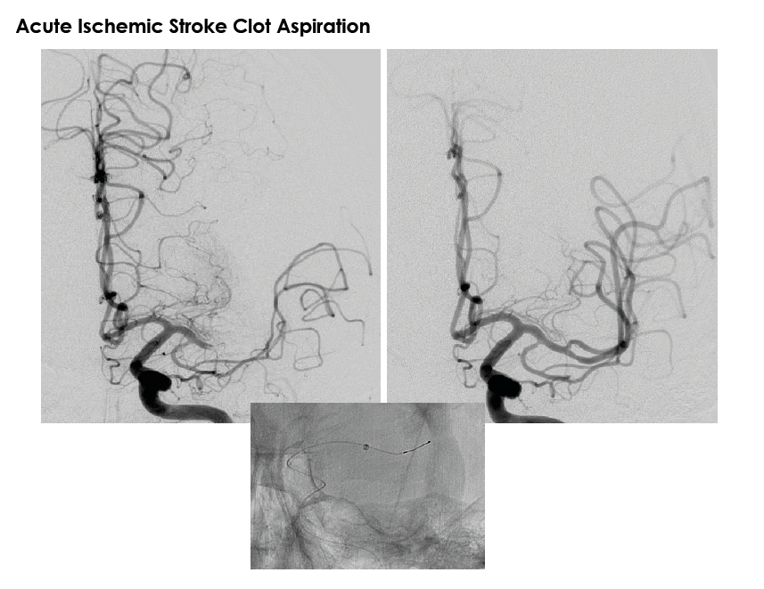
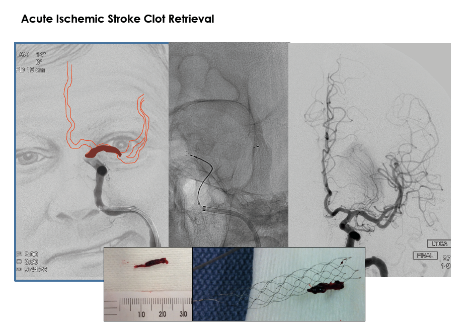
The impact of ischemic stroke upon healthcare in the United States is staggering. Extensive research has attempted to unlock methods of safely restoring blood flow to the brain and protect patients from progression to permanent brain damage. In 1996, the United States Food and Drug Administration approved the use of intravenous tissue plasminogen activator (IV tPA) for use in the treatment of acute ischemic stroke in selected patients. This “clot-busting” medicine has been shown to improve patient outcomes when it is administered within the first few hours of a stroke. Among those patients who are not candidates for an intravenous medication, some may benefit from an endovascular method of removing a blood clot. Endovascular methods include navigating a small catheter (a hollow plastic tube) through the blood vessels to the location of the blockage and using one of several therapies. After a catheter is positioned within the blocked artery, tPA can be injected to dissolve the clot, or a device can be used to suction the clot (aspiration) or trap the clot and remove it from the body. Time is brain, which is why methods of opening a blocked artery are performed in the first few hours of an ischemic stroke.

Acute Ischemic Stroke Clot Aspiration

Acute Ischemic Stroke Clot Retrieval

Epistaxis Embolization Therapy
Epistaxis, or excessive bleeding from the nose, can be caused by various underlying conditions. In cases of severe bleeding that is not controlled with conventional surgical techniques, endovascular embolization therapy can be performed. In this procedure, a small catheter is advanced under x-ray guidance to the arteries that serve the nose. Small particles are then injected to intentionally plug the arteries and stop the bleeding.

Inferior Petrosal Sinus Sampling
Cushing syndrome results from excess production of the hormone cortisol. The release of cortisol is controlled by adrenocorticotropic hormone (ACTH), which can be produced in several locations throughout the body. One location is the pituitary gland, which is located in a region behind the eyes and inside the skull. An evaluation for a patient with Cushing syndrome may include inferior petrosal sinus sampling (IPSS). In this procedure, two microcatheters are navigated under x-ray guidance to venous sinuses in the region of the skull base deep behind the nose, the location where the pituitary gland drains. A medication (corticorelin) is then administered and serial blood samples are obtained and tested for levels of hormone to determine the location of abnormal ACTH release.

Inferior Petrosal Sinus Sampling
Intracranial Balloon Angioplasty and Stenting
Ischemic stroke can have multiple causes including clot migration from the heart, narrowing of arteries in the neck, and plaque buildup leading to narrowing of the blood vessels in the brain, which is known as intracranial atherosclerotic disease. Management for patients who have had a stroke due to intracranial atherosclerotic disease includes aggressive medical management with vigilant control of risk factors such as hypertension, diabetes and elevated cholesterol, as well as smoking cessation and antiplatelet medicines. Some patients may be candidates for additional therapies known as intracranial balloon angioplasty or stenting. Balloon angioplasty utilizes endovascular techniques to access the inside of the blood vessel at the region of narrowing, where a balloon is temporarily inflated to expand the narrowed region and restore normal blood flow. A similar technique involves the additional placement of a stent within the blood vessel at the region of narrowing.

Intracranial Balloon Angioplasty and Stenting
Intracranial Venous Sinus Stenting

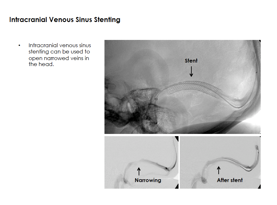
Intracranial venous sinus abnormalities can obstruct the outflow of blood from the head. In some patients who have chronically elevated intracranial pressures, venous outflow obstruction may be a related finding. For evaluation, patients often undergo diagnostic brain imaging such as MRI or MR angiography, detailed eye examination, and an endovascular procedure to perform manometry testing to evaluate intracranial pressures in several locations. Selected patients may benefit from placement of a stent within a dural sinus of the head to improve venous outflow. The stent is delivered using standard neuroendovascular techniques and on occasion a balloon is also inflated within the stent to ensure optimal placement.

Intracranial Venous Sinus Stenting

Preoperative Tumor Embolization
Tumors can be highly vascularized structures and this large amount of blood vessels can lead to excessive bleeding during surgical removal. In some cases, patients undergoing a surgical resection of a tumor of the head, neck, or spine are referred for a procedure to reduce blood supply to the tumor. This procedure is known as pre-operative tumor embolization. A small catheter is advanced under x-ray guidance into the region of the blood vessels supplying a tumor. After angiographic evaluation of the blood supply, blood vessels can be intentionally plugged by injecting small particles or glue-like material into the arteries. This technique aims to “devascularize,” the tumor and reduce intra-operative bleeding.

Preoperative Embolization of Head, Neck, and Spine Tumors
Vertebral Artery Balloon Angioplasty Stenting
Stenosis at the origin of the vertebral artery is a recognized risk factor for ischemic stroke. Patients who have symptoms of stroke or transient ischemic attack (TIA) may undergo diagnostic imaging to evaluate the blood vessels of the head and neck. Patients with severe stenosis at the origin of the vertebral artery may benefit from endovascular treatment. After a catheter is navigated to the area of narrowing, a small wire is advanced through the region of stenosis and positioned in the vertebral artery. A balloon-mounted stent is typically used, which is advanced over the wire to the area of narrowing and the balloon is inflated to both expand the narrowing and implant the stent. Additional balloon inflations can be performed before and after the stent placement if needed to ensure adequate expansion of the narrowed region. This procedure is commonly performed with temporary placement of a filter downstream from the region of plaque to capture any debris that may become dislodged during the stent placement.

Vertebral Artery Balloon Angioplasty and Stenting
Wada Testing
A Wada test is used for pre-operative surgical planning in epilepsy. Patients who have epilepsy that is refractory to medications, and who are selected as candidates for surgical resection of a region of abnormal brain tissue, may undergo a Wada test. The test is performed in the angiography suite, similar to all other neurointerventional procedures. Electroencephalogram (EEG) leads are placed on the head to measure brain wave recordings and a diagnostic catheter cerebral angiogram is performed to evaluate the intracranial blood supply. A medication (sodium amytal) is then injected into a blood vessel to temporarily put half of the brain to sleep. The patient is then engaged in a series of memory and language tests. The medication wears off in a few minutes and the procedure is then repeated for the other half of the brain. This test is intended to determine which side of the brain controls language function and identify the role that each hemisphere plays in memory.

Disclaimer

The information on the Division of Stroke, Neurointervention and Critical Care website is for educational purposes only and is not intended as medical advice. While general information regarding the conditions treated and procedures performed is posted, this information is not a substitute for professional medical advice, diagnosis, or treatment. All patients are treated with individualized care based on independent professional judgment and experience in each specific case.