
Oxford Instruments Center for Advanced Microscopy – Electron Microscopy (OxCAM-EM) Core
The OxCAM-EM Core is an institutionally available research service unit managed on behalf of the Medical College by the Department of Cell Biology, Neurobiology and Anatomy. The Core encompasses both Light and Electron Microscopy services and is located on the 4th Floor of the MACC Fund Building. OxCAM-EM is open to all Medical College of Wisconsin faculty, staff, and students; investigators at affiliated institutions; and other non-MCW investigators. The Core operates on a fee-for-service basis.
Front: Megan Harwig, Melissa Niehausen, Lorenzo Bacalzo
Back: Ashish Gadicherla, Rob Goodwin View schedules and rates in iLab
Front: Megan Harwig, Melissa Niehausen, Lorenzo Bacalzo
Back: Ashish Gadicherla, Rob Goodwin View schedules and rates in iLab

OxCAM-EM Core Featured Equipment
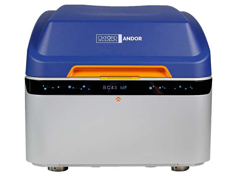
BC43
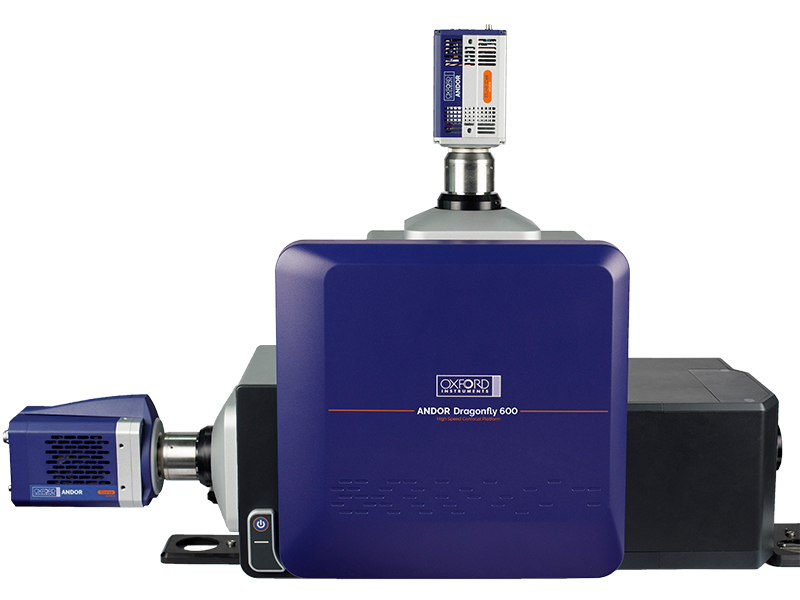
Dragonfly 600
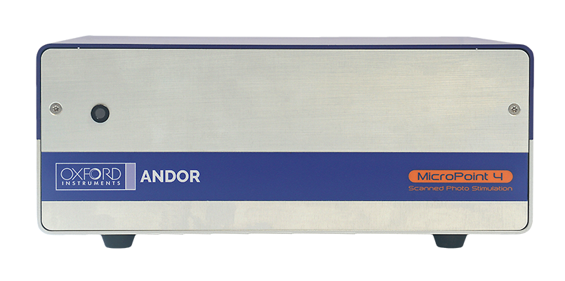
MicroPoint 4
Optical Microscopy Resources
The Core relies primarily on a teach-and-train model for these microscopes.
In addition to these microscopes, the Core also has facilities for image processing using Imaris software packages.
Andor BC43
Andor Dragonfly 502
Andor Dragonfly 620SR
Aberrior Facility Line
Nikon N-SIM
Electron Microscopy Resources
The Electron Microscopy Facility provides service, consultation for research, and some training for projects requiring transmission electron microscopy.
Jeol 1400Flash Transmission Electron Microscope
Leica EMPact 2 High Pressure Freezing Apparatus
RMC PowerTome Ultramicrotomes
Prepmaster Specimen Preparation Robot
Quorum Technologies 150T E Sputter Coater/Turbo Evaporator (EMS 150T E)
PELCO easiGlow Glow Discharge System
Meet Our Team

Ashish Gadicherla, PhD
Assistant Professor, Director, Oxford Instruments Center for Advanced Microscopy, EM Core Facility Technical Director, Structural Biology Shared Resources, MCW Cancer Center


Lorenzo Bacalzo
Research Technologist II

Robert Goodwin
EM Core Lab Manager

Megan Harwig
Research Scientist II; Light Microscopy Manager
