
Medical College of Wisconsin Neuro-Oncology Brain Bank and Associated Research
Dr. Jennifer Connelly is actively involved in ongoing projects led by Dr. Peter LaViolette’s lab in the Department of Radiology. Ongoing efforts are focused on creating and validating imaging techniques that are meant to improve both patient outcome and treatment efficacy. Our lab primarily works with brain cancer patients in the translational setting, and our ongoing collaborative projects include additional clinical members of neurology, neuro-oncology, neurosurgery, radiology, pathology, and radiation oncology. Our primary goal is to make our imaging technology more sensitive and specific for detecting brain cancer growth and response to treatment.
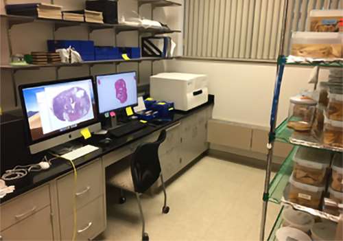
Pictured right: The tissue bank in the LaViolette Lab and Neuro-Oncology Brain Bank. The Huron slide scanner is on the bench next to a Windows work station equipped with Huron software. The iMac workstation shown is a hub for a 70Tb drive, which stores the raw slide data. On the right are whole brain samples from the Neuro-Oncology Brain Bank.
Research Areas
Magnetic Resonance Imaging
Patients being treated for brain cancer undergo MRI scans repeatedly during treatment. The imaging studies allow radiologists to determine whether tumors are growing or remaining stable. While the technology driving the machines is relatively mature, brain tumors often invade well beyond the margins detectable with today’s imaging.
Brain Bank
Validation of imaging requires a ‘ground truth’ comparison with known tissue types. Because of this limitation, and due to the fact that brain tumors change during the course of treatment, whole brain samples are ideal. In 2010, Drs. Connelly, Cochran, and LaViolette enrolled the first patient in our brain donation program. Since then, our recruitment has been formalized and a formal tissue bank has been formed. Patient recruitment has steadily increased, driven largely by Dr. Connelly. To the best of our knowledge, our brain bank is the largest brain cancer specific whole brain tissue bank in the world. We have recruited over 65 patients for participation, and many brain tumor types and treatment histories are represented.
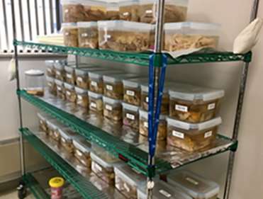
Pictured left: The Neuro-Oncology Brain Bank is located on the 4th floor of the MACC Fund Research Building and currently houses over 65 whole brain samples from brain cancer patients and is the largest such resource in the country.
Unique Equipment and Database
The Neuro-Oncology Brain Bank lab also houses a rapidly expanding database that contains both the clinical imaging from each patient, clinical histories, and digital histology obtained from each case. This resource is housed locally within the lab on servers with over 120Tb of storage. Additional MCW resources such as the Research Computing Center (RCC) are also utilized for high-throughput computing and long-term storage. As this database grows, future resources will be necessary to continue to support it, which will include additional storage, and additional high-performance computer systems.
3D Printers
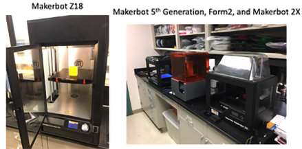
The Neuro-Oncology Brain Bank is equipped with four 3D printers, three Makerbots and one Form Labs Form2. The Makerbots are used to print the prostate and brain slicing jigs used for aligning pathology with imaging. We also 3D print models for surgical planning purposes.
Slide Digitization Microscope
The LaViolette Lab recently purchased a Huron slide scanner capable of scanning three large format histology slides at once at 40X resolution. This resource is located in the tissue bank room and is being used for digitizing both H&E stained slides and immunohistochemistry slides for molecular markers.
Meet Our Team

Jennifer M. Connelly, MD
Professor
Peter S. LaViolette, PhD, MS
Robert C. Olson, MD, Professorship in Radiology; Director, Quantitative Imaging Laboratory, Medical College of Wisconsin; Section of Imaging Research, Division of Imaging Sciences
L. Tugan Muftuler, PhD
Professor

Andrew S. Nencka, PhD
Professor; Director, Center for Imaging Research (CIR); Section of Imaging Research, Division of Imaging Sciences
Funding Sources & Active Grants
The LaViolette Lab and Neuro-Oncology Brain Bank is currently funded by the National Institute of Health (NIH), the National Cancer Institute (NCI), through an academic-industrial partnership with Novocure Inc. and through a pilot grant funded by philanthropic funds earmarked for neurooncology research at Froedtert and MCW. Past grants have come from the MCW Research Affairs Committee, the American Brain Tumor Association, and the American College of Radiology Imaging Network.
Active Grants
Brain Cancer Radio-Pathomics for Predicting Heterogeneous Cytology
Source:: NIH/NCI R01CA218144
Key Personnel: LaViolette (PI), Connelly, Cochran, Nencka, Muftuler, Banerjee (Co-Investigators).
Dates: 7/1/2017 – 6/30/2022
Funds: $1,752,950 (total for all years)
Alzheimer’s Disease Radio-Pathomics for Predicting Heterogeneous Cytology
Source: NIH/NCI R01CA218144-02S1
Key Personnel: LaViolette (PI), Connelly, Cochran, Nencka, and Brehler (Co-Investigators).
Dates: 6/1/2018 – 5/31/2021
Funds: $192,500 (total for all years)
NovoTTF Treatment Signatures in Glioblastoma Patients at Autopsy
Source:: Novocure Inc.
Key Personnel: LaViolette (PI), Connelly, Cochran (Co-Investigators)
Dates: 06/01/2017 - 05/30/2021
Funds: $294,451 (total for all years)
Imaging signatures of TTField Treatment at Autopsy
Source:: Novocure Inc.
Key Personnel: LaViolette (PI), Connelly, Cochran (Co-Investigators)
Dates: 01/01/2019 - 05/30/2021
Funds: $115,000 (total for all years)
The molecular evolution of gliomas, from diagnosis to end of life.
Source: Froedtert Foundation and Neuro-oncology Philanthropy
Key Personnel: LaViolette (PI), Connelly, Cochran (Co-Investigators), Roel Verhaak (Jackson Laboratory).
Dates: 1/1/2019 - 012/31/2021
Funds: $31,874 (total for all years)
Current Projects
Our research projects are funded by the National Institute of Health (NIH), the National Cancer Institute (NCI), and through an academic-industrial partnership with Novocure Inc.