
About the Yoganandan Laboratory
The Yoganandan Laboratory conducts transformational research studies in the field of biomechanics and neuroscience. Ongoing research utilizes computer and statistical models to develop personalized medicine for spinal disorders, develop response corridors and injury risk functions for the head and spine, and determine human head-spine responses under physiologic and traumatic loads.
Research Areas
Personalized Medicine for Cervical Spine Degeneration and Deformity Surgery
The Yoganandan Laboratory uses head-to-spine models to study the effectiveness of different surgical procedures. Head-to-spine studies include classical anterior cervical discectomy and fusion (ACDF) and cervical disc arthroplasty (CDA). This research examines the role of CDA and ACDF on indexical and adjacent levels for clinical metrics, such as range of motion, and biomedical metrics, such as disc and facet load sharing. Areas of focus include heterotopic ossification, an unintended consequence of CDA, and accelerated adjacent-segment degeneration, which may lead to additional surgeries. Both single and contiguous two-level and hybrid options are being evaluated under physiological and traumatic forces. Our gender-specific computer finite element models of the head and neck with musculature can be morphed to mimic each patient’s anatomy. This allows us to depict the disorder/abnormal situation using MRI, elucidate different surgical options (CDAs and ACDFs), and simulate long-term spinal changes. These multimodal personalized medicine models can be used for patient education, and patient-specific models serve as another surgical decision-making tool for optimum selection and treatment of patients with cervical spine disc diseases such as degenerative spondylotic myelopathy (DCM).
Funding
This work is part of a CDMRP study funded by the US Department of Defense.
Representative Publications
- Biomechanical effects of uncinate process excision in cervical disc arthroplasty
- Effect of heterotopic ossification after Bryan-cervical disc arthroplasty on adjacent level range of motion: A finite element study
- Comparison Study of Four Cervical Disk Arthroplasty Devices Using Finite Element Models.
- Influence of Cervical spine sagittal alignment on range of motion after corpectomy; a finite element study.
- Comparative Finite Element Modeling Study of Anterior Cervical Arthrodesis Versus Cervical Arthroplasty with Bryan Disc or Prodisc C.
Image Description
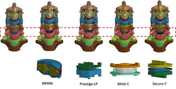
The included image depicts a finite element model of intact spine, coronal view (top left); finite element model of spine with implantation of Bryan implant, Prestige-LP implant, Mobi-C implant, Secure-C implant (top center left to far right); finite element model of Bryan implant, Prestige-LP implant, Mobi-C implant and Secure-C implant (bottom left to right).
Personalized Medicine for Lumbar Spine Degeneration and Deformity Surgery
Following the concept of the head-neck study, a series of morphable lumbar spine-pelvis finite element models is being developed to study degenerative diseases and surgical options for the low back. Procedures such as anterior lumbar interbody fusion (ALIF), posterior lumbar interbody fusion (PLIF), transforaminal lumbar interbody fusion (TLIF), oblique lateral interbody fusion (OLIF), and eXtreme lumbar interbody fusion (XLIF) with physiological biomechanical loads on each patient are simulated using finite element models. As above, this study is examining the role of different types of procedures on index and adjacent levels for clinical outcomes. Our computer models are morphable to mimic each patient’s anatomy depicting the abnormal/diseased spine using routine medical imaging (MRI), simulate models with different surgical options, and simulate long-term spinal changes. These personalized medicine models are useful for patient education and act as a surgical decision-making tool for optimum treatment of patients with lumbar spine disease.
Funding
This model is being used in the cited CDMRP grant for determining human tolerance to injury from events such as underbody blast and the role of body armor on spinal loading.
Representative Publications
- Development and validation of osteoligamentous lumbar spine under complex loading conditions: A step towards patient-specific modeling
- Biomechanical investigation of lumbar interbody fusion techniques.
- Importance of neural foraminal narrowing in lumbar spine fractures of low AIS severity.
Image Description
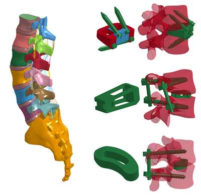
The included image shows intact lumbosacral spinal column (left) and models with different surgical options, including ALIF, PLIF and TLIF (right, top to bottom).

Biomedical Models and Neurotrauma
The Yoganandan Laboratory is collaborating with other investigators in the Zablocki VA Medical Center Laboratories on a series of studies delineating injury mechanisms and determining human tolerances. These studies are aimed at improving safety in real-world traumatic events, developing standardized testing methodologies, and establishing federal standards. They incorporate experimental models using pathological specimens, computerized whole-human body models, computerized regional models for areas such as head and spine, statistical risk analysis models, and field database analyses. Currently funded studies are focused on lumbar spine-pelvis injuries from vertical loading for automotive, military, aviation, and other applications.
Funding
This project is part of a series of studies on head and spine (and other body regions) for the US Department of Transporation (DOT), US Department of Defense (DOD), and others.
Representative Publications
- Uncertainty Evaluations for Risk Assessment in Impact Injuries and Implications for Clinical Practice.
- Human Pelvis Injury Risk Curves from Underbody Blast Impact.
- Human Lumbar Spine Responses from Vertical Loading: Ranking of Forces Via Brier Score Metrics and Injury Risk Curves.
- Role of age and injury mechanism on cervical spine injury tolerance from head contact loading.
- Role of disc area and trabecular bone density on lumbar spinal column fracture risk curves under vertical impact.
- Upright Magnetic Resonance Imaging Study of Cervical Flexor/Extensor Musculature and Cervical Lordosis in Females After Helmet Wear.
Image Descriptions
Top image: Top row shows Coronal CT scan pre-test (left), coronal CT scan post-test showing injuries at the upper lumbar vertebrae (middle), and post-test photograph of the specimen in the coronal plane showing injuries, although to a less demonstrable extent, at the inferior level (right). Bottom row shows pre-test sagittal CT scan (left), post-test sagittal CT (middle), and post-test photograph of the specimen in the sagittal plane (right). Comparison demonstrates that CT images are more effective in showing the pathology in both planes.
Bottom image: Survival analysis-based injury risk curve with ±95% confidence intervals (dashed line) for lumbar spine fracture under axial loading.

Shared Facilities at ZVAMC Labs
Located on the Zablocki VA Medical Center Campus, the Yoganandan Laboratory enjoys the use of shared facilities specifically designed to support investigations into tissue and spine biomechanics, as well as related computational modeling and statistical validation.
People
Led by Dr. Narayan Yoganandan, the Yoganandan Lab works closely with internal collaborators from the Medical College of Wisconsin and shared engineering and research staff of the Zablocki VA Medical Center Laboratories. Meet just a few of these key individuals below.

Narayan Yoganandan, PhD
Professor & Chair of Biomechanics, Department of Neurosurgery; Orthopaedic Surgery; Joint Department of Biomedical Engineering

Matthew D. Budde, PhD
Associate Professor

Frank A. Pintar, PhD
Professor
Alok Shah
Director of Biomechanics Research
Brian D. Stemper, PhD
Professor, Joint Department of Biomedical Engineering; Neurosurgery

Aditya Vedantam, MD
Associate Professor; Director, Minimally Invasive Surgery; Director, Center for Cervical Myelopathy; Adjunct Faculty in Biomedical Engineering, Radiology, and Orthopedics
Recent Publications
-
(Seifert J, Koser J, Shah A, Frazer L, Pintar FA, Yoganandan N, Nicolella D, Bentley TB, Stemper BD.) Ann Biomed Eng. 2026 Apr;54(4):1117-1125 PMID: 40658298 SCOPUS ID: 2-s2.0-105010678874 07/14/2025
-
Mechanical Characterization of Non-degraded Porcine Annulus Fibrosus Material Properties.
(Seifert J, Maiman D, Frazer LL, Shah A, Yoganandan N, King K, Sheehy JB, Paskoff G, Bentley T, Nicolella DP, Stemper BD.) Ann Biomed Eng. 2026 Apr;54(4):1099-1107 PMID: 39499364 SCOPUS ID: 2-s2.0-85208479174 11/05/2024
-
(Kote VB, Frazer LL, Hostetler ZS, Jones DA, Davis M, Op’t Eynde J, Kait J, Pang D, Bass D, Koser J, Shah A, Yoganandan N, Stemper B, Bentley T, Nicolella DP.) Annals of Biomedical Engineering. April 2026;54(4):1023-1037 SCOPUS ID: 2-s2.0-85197253915 04/01/2026
-
Lumbar Spine Orientation Affects Compressive Fracture Outcome
(Cutlan R, Khokhar M, Shammout N, Shah AS, Frazer L, Yoganandan N, Shender BS, Sheehy J, Paskoff G, Nicolella D, Bentley T, Shabani S, Stemper BD.) Annals of Biomedical Engineering. April 2026;54(4):1076-1085 SCOPUS ID: 2-s2.0-85207724941 04/01/2026
-
(Banerjee A, Hu H, Yoganandan N.) Journal of the Indian Society for Probability and Statistics. December 2025;26(2):631-652 SCOPUS ID: 2-s2.0-105007001437 12/01/2025
-
(Rahman M, Banurekha Devaraj K, Chauhan O, Harinathan B, Yoganandan N, Vedantam A.) Applied Sciences Switzerland. January 2025;15(2) SCOPUS ID: 2-s2.0-85215797547 01/01/2025
-
Expanded Combined Loading Injury Criterion for the Human Lumbar Spine Under Dynamic Compression.
(Ortiz-Paparoni M, Op 't Eynde J, Eckersley C, Morino C, Abrams M, Pang D, Kait J, Pintar F, Yoganandan N, Moore J, Barnes D, Loftis K, Bass CR.) Ann Biomed Eng. 2024 Nov;52(11):3067-3077 PMID: 39240473 SCOPUS ID: 2-s2.0-85203121909 09/06/2024
-
(Bakhaidar M, Harinathan B, Devaraj KB, DeGroot A, Yoganandan N, Shabani S.) Int J Spine Surg. 2024 Sep 12;18(4):441-447 PMID: 39214603 PMCID: PMC11483624 08/31/2024
-
Behind Armor Blunt Trauma: Liver Injuries Using a Live Animal Model.
(Yoganandan N, Shah A, Koser J, Stemper BD, Somberg L, Chancey VC, McEntire J.) Mil Med. 2024 Aug 19;189(Suppl 3):659-664 PMID: 39160873 SCOPUS ID: 2-s2.0-85201736818 08/20/2024
-
Is Posterior Cervical Foraminotomy Better Than Fusion for Warfighters?: A Biomechanical Study.
(Choi H, Purushothaman Y, Ozobu I, Yoganandan N.) Mil Med. 2024 Aug 19;189(Suppl 3):710-718 PMID: 39160815 SCOPUS ID: 2-s2.0-85201744350 08/20/2024
-
Hybrid III Manikin Lumbar Spine Loading Under Vertical Impact.
(Yoganandan N, Moore J, Westerhof TA, Flierman NA.) Mil Med. 2024 Aug 19;189(Suppl 3):55-62 PMID: 39160828 SCOPUS ID: 2-s2.0-85201737711 08/20/2024
-
(Rahman M, Palmer P, Harinathan B, Banurekha Devaraj K, Yoganandan N, Vedantam A.) Diagnostics (Basel). 2024 Jul 12;14(14) PMID: 39061634 PMCID: PMC11276270 07/27/2024
Educational Opportunities in Biomedical Engineering
Get Involved
Graduate Education
Researchers seeking Graduate-level opportunities at the cross-section of Trauma Biomechanics and Neuroscience are invited to explore opportunities in the Marquette-MCW Joint Department of Biomedical Engineering, as well as the Neuroscience Doctoral Program at the Medical College of Wisconsin.
Student Research
Undergraduate and high school students interested in gaining research experience in neuroscience and biomedical engineering labs are invited to explore undergraduate research opportunities supported by the Zablocki VA Medical Center Laboratories.
ZVAMC Educational Opportunities
General Inquiries
For general inquiries or to learn more about employment or ongoing research, please contact ZVAMC Labs Project Coordinator, Christy Stadig.