Case Study 18 - CC: 8-year-old boy presents with difficulty seeing whiteboard at school
Original Authors: Polina Prokhoda, Albert Kim, MD, Joseph Carroll, PhD
HPI:
An 8-year-old male patient presents with mother of child to general ophthalmology clinic. Patient and mother served as independent historians. He has not been seen by an ophthalmologist or optometrist before but has been having vision checks at school and sees his pediatrician every couple of years. Mother reports that for the last 6 months her son has been complaining of difficulty seeing the whiteboard at school. Patient reports that the light sometimes makes his eyes water. Mom has not noticed any eye crossing or eye wandering, but does note his eyes move back and forth at times, worse when he is tired. According to mom, other than noting difficulty seeing the board at school, patient’s vision does not seem to prevents him from playing with friends or doing any other activities.
Past Ocular History:
None
Ocular Medications:
None
Past Medical History:
Born at 39 weeks without complications
Frequent ear infections prior to ear tubes
Surgical History:
Bilateral ear tubes at age 2
Past Family Ocular History:
No history of blinding diseases, glaucoma or macular degeneration
Social History:
Lives at home with mom and dad. No smoker in home. Currently in 3rd grade.
Medications:
None
Allergies:
No known drug allergies
ROS:
Denies recent illness or any new CNS, heart, lung, GI, skin or joint symptoms.
Ocular Exam
Visual Acuity (cc):
OD: 20/50
OS: 20/50
Visual acuity OU improves to 20/40 with pinhole and refraction
IOP (iCare tonometry):
OD: 16 mmHg
OS: 17 mmHg
Pupils:
OD: Equal, round and reactive to light in dark and light settings, no APD
OS: Equal, round and reactive to light in dark and light settings, no APD
Extraocular Movements:
OD: Full. Mild nystagmus noted at distance and at near.
OS: Full. Mild nystagmus noted at distance and at near.
No anomalous head posture or strabismus noted.
Confrontational Visual Fields:
OD: Full to finger counting
OS: Full to finger counting
Slit Lamp Exam:
| RIGHT EYE | LEFT EYE | |
| External | Normal |
Normal |
| Lids and Lashes | Normal | Normal |
| Conjunctiva/Sclera | White and quiet | White and quiet |
| Cornea | Clear |
Clear |
| Anterior Chamber | Deep and quiet | Deep and quiet |
| Iris | Normal | Normal |
| Lens | Clear | Clear |
| Anterior Vitreous | Normal | Normal |
| RIGHT EYE | LEFT EYE | |
| Disc | Sharp optic disc margins | Sharp optic disc margins |
| C/D Ratio | 0.2 with 90D lens | 0.2 with 90D lens |
| Macula | Blunted foveal light reflex | Blunted foveal light reflex |
| Vessels | Normal | Normal |
| Periphery | Blonde fundus | Blonde fundus |
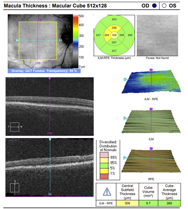
OCT Imaging:
Image demonstrates lack of normal foveal contour due to foveal hypoplasia
Fundus Photo:
Image demonstrates lack of foveal light reflex and blonde fundus.
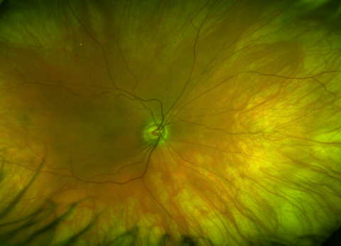
Image references: Patient JC_12565 seen at AOIP with known ocular albinism diagnosis.
Case Studies
-
Ophthalmic Case Study 1
Acute right eye pain -
Ophthalmic Case Study 10
Blurry vision in the left eye for 2 weeks -
Ophthalmic Case Study 2
Red, itchy eyes -
Ophthalmic Case Study 11
Acute pain and burning in L eye -
Ophthalmic Case Study 3
Acute left eye pain and blurry vision -
Ophthalmic Case Study 12
Blurry vision in both eyes and headaches -
Ophthalmic Case Study 4
Left eye pain and fuzzy vision 2 days after eye surgery -
Ophthalmic Case Study 13
"Cannot see well" from left eye -
Ophthalmic Case Study 5
Girl rubbing her R eye after trauma -
Ophthalmic Case Study 14
Blurry vision in both eyes -
Ophthalmic Case Study 6
Red eye and pain on the left -
Ophthalmic Case Study 15
Eye irritation and dryness -
Ophthalmic Case Study 7
Vision loss L eye -
Ophthalmic Case Study 16
2 brief episodes of vision loss in the R eye -
Ophthalmic Case Study 8
Crossed eyes -
Ophthalmic Case Study 17
Routine eye exam -
Ophthalmic Case Study 9
White pupils
Contact Ophthalmology
For patient care inquires, call us at (414) 955-2020 or use MyChart. Email is for research and education inquiries only.
Eye Institute Location
925 N. 87th St.
Milwaukee, WI 53226
Appointments
(414) 955-2020
Continuing Medical Education
Renee Ware
(414) 955-2086
Medical Education Coordinator
Associate Director of Development - Ophthalmology
Sarah Walker
Refer to Us - Consultation requests
Patient Referral Form (PDF)
Fax to (414) 955-0136
Emergent Requests
Within 48 hours call
(414) 955-2020
Research
Vesper Williams
(414) 955-7862
Advanced Ocular Imaging Program
(414) 955-2647
Eye Institute Executive Director (Administrator)
Shannon Dreier