header-logo
Ophthalmology_Hero Image 2

Case Study 6 - CC: Red eye and pain on the left

all
Patient Visit

Patient History
HPI:

42 year-old African American male policeman presents to clinic reporting a sore and red eye shortly after a being elbowed during a drug dealer sting operation. He was not wearing protective eyewear. He reports blurry vision. He reports tenderness over his left eye, eyebrow, and cheekbone.

Past Ocular History:
None

Ocular Medications:
None

Past Medical History:
Sickle cell trait, type 2 diabetes mellitus, hypertension

Surgical History:
None

Past Family Ocular History:
No family history of glaucoma, macular degeneration or other blinding diseases

Social History:
Drinks socially, no cigarette or illicit drug use.

Medications:
Metformin
Glipizide
Aspirin 81mg
Hydrochlorothiazide

Allergies:
None

ROS:
No known chronic inflammatory disease. No history of easy bleeding or bruising. No recent illnesses or other CNS, heart, chest, GI, joint or skin problems.

Ocular Exam

Visual Acuity (cc):
OD: 20/20
OS: 20/40

IOP (tonoapplantation):
OD: 15 mmHg
OS: 23 mmHg

Pupils:
Normal pupil reaction R, sluggish L pupil, no APD.

Extraocular Movements:
Full OU. No nystagmus.

Confrontational Visual Fields:
Full to finger counting OU

External:
Normal R; moderate upper lid edema and mild erythema L

Slit Lamp:

Lids and Lashes Normal OU
Conjunctiva/Sclera Normal OD, mild conjunctival hyperemia 360 degrees OS
Cornea Normal OU, no corneal epithelial defects
Anterior Chamber Deep and quiet OD, hyphema obscuring 1/3 of the anterior chamber OS
Iris Normal OU, no obvious neovascularization OU
Lens Normal OU, no evidence of cataract or dislocated lens OS
Anterior Vitreous Clear OU

Dilated Fundus Examination:
OD Clear view; CDR: 0.3 with sharp disc margins; flat macula with normal red reflex; normal retina vessels and periphery.
OS Slight hazy view; CDR: 0.3; no obvious vitreal heme, retinal tear or detachment or choroidal rupture
Other:
CT orbit - round globe. No intraorbital pathology. Mild upper lid edema.
Diagnosis and Discussion

Diagnosis
Traumatic hyphema

Discussion

Differential Diagnosis: This patient’s clinical presentation and exam findings are consistent with the diagnosis of traumatic hyphema. In cases of non-traumatic hyphema diagnoses to consider include: iris neovascularization, herpes simplex/zoster iridocyclitis, hx of blood dyscrasias and intraocular tumors.

Definition:
Hyphema is defined as blood in the anterior chamber. When enough blood accumulates, the blood precipitates in the inferior angle covering anterior segment structures. Hyphemas are classified as followed: grade I hyphema- less than 1/3 of the inferior anterior chamber is covered with blood, grade II- 1/3 to 1/2 of the anterior chamber, grade III-1/2 to nearly total of the anterior chamber, grade IV- total hyphema (if black in color named 8-ball hyphema). Bleeding usually occurs from the rupture of ciliary vessels.

Examination:
Work-up for hyphema should include a detailed history on the type of injury, personal or family history of sickle cell disease, use of anticoagulants, and history of coagulopathy. On exam, FIRST RULE OUT AN OPEN GLOBE. Other important aspects of the exam include documentation of the extent and location of the hyphema, measurement of IOP, and DFE without scleral depression. CT of the orbits can be performed if indicated as in cases of facial/orbital fractures. A B-scan ultrasound can be considered if there is poor visualization of the posterior pole. African American and Mediterranean patients should be screened for sickle cell disease as they are at risk for increased IOP and vision loss due to trabecular meshwork obstruction by the sickled cells.

Treatment:
In the treatment of hyphema, it is crucial to stress the importance of complete bed rest. Additionally, patients should be instructed to elevate the head of the bed to allow the blood to settle inferiorly as not to obstruct the visual axis. A shield should be placed over the involved eye at all times to prevent further trauma. Non-NSAID analgesics should be used for pain relief. Cycloplegics (such as atropine or scopolamine) and topical steroids are recommended to treat the associated iritis and prevent synechiae. If patients have increased IOP, first line therapy is topical beta-blocker but additional medical therapy should be added to maintain normal IOP. Indications for surgical evaluation include: corneal stromal blood staining, non-clearing total hyphema and high IOP. The threshold for surgical evacuation is lower in sickle cell patients due to a higher risk of permanent vision loss.

Patients should be followed frequently in the first 2 weeks to monitor IOP and treat any other pathology. The risk of rebleeding is highest in the 3 – 7 day period post injury and is associated with worse prognosis. Complications of hyphema include posterior synechiae, peripheral anterior synechiae, corneal blood staining, glaucoma and optic atrophy due to high IOP. Prognosis can be poor to excellent depending on the severity of initial injury. Patients should be followed annually to monitor for development of glaucoma and other pathology.

Self-Assessment Questions
  1. In patients with a history of trauma and blood in the anterior chamber what needs to be ruled out?
  2. What laboratory test would you order in a patient with a hyphema and African American or Mediterranean heritage?

References/Resources

Self-Assessment Answers

In patients with a history of trauma and blood in the anterior chamber what needs to be ruled out?

d. evidence of a ruptured globe

A comprehensive exam should be performed to ensure no globe rupture.

What laboratory test would you order in a patient with a hyphema and African American or Mediterranean heritage?

a. Sickle cell test

Patients with sickle cell trait or sickle cell anemia might develop high intraocular pressure due to blood in the anterior chamber and are at increased risk of optic nerve head damage and vision loss.

Contact Ophthalmology

For patient care inquires, call us at (414) 955-2020 or use MyChart. Email is for research and education inquiries only.

Eye Institute Location

925 N. 87th St.

Milwaukee, WI 53226


Appointments

(414) 955-2020

(414) 955-6166 (fax)


Continuing Medical Education

Amanda Tan

atan@mcw.edu

(414) 955-2049


Medical Education Coordinator

Ophth-Residency@mcw.edu


Associate Director of Development - Ophthalmology

Sarah Walker

sarawalker@mcw.edu

Refer to Us - Consultation requests

Patient Referral Form (PDF)

Fax to (414) 955-0136


Emergent Requests

Within 48 hours call

(414) 955-2020


Research

Vesper Williams

vewilliams@mcw.edu

(414) 955-7862


Advanced Ocular Imaging Program

aoip@mcw.edu

(414) 955-2647


Eye Institute Executive Director (Administrator)

Shannon Dreier
sdreier@mcw.edu

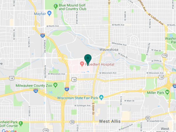
Eye Institute Google map location