Case Study 1 - CC: Acute R Eye Pain
Diagnosis
Suspected acute angle closure glaucoma R eye
Narrow angle L eye
Discussion
 Differential Diagnosis:
The patient described is presenting with acute angle closure glaucoma due to shallow anterior chamber angles. Other causes of increase intraocular pressure include open angle glaucoma or other pathology of the trabecular meshwork (pigmentary dispersion, pseudoexfoliation, trauma, neovascularization, etc.) that obstructs aqueous outflow. Forward displacement of intraocular structures (posterior tumor, choroidal swelling, etc.) can also block the outflow of aqueous and increased the intraocular pressure.
Definition:
Acute angle closure glaucoma occurs when there is a relatively sudden blockage of the trabecular meshwork causing elevation of the intraocular pressure. One possible mechanism is the anterior bulging of the peripheral iris, occluding the trabecular meshwork and trapping aqueous humor inside the eye. The reason for the sudden anterior bulging of the peripheral iris may be due to pupillary block (where the lens presses up against the iris trapping all the aqueous humor behind it). This results in increased fluid in the posterior chamber, creating a pressure gradient that subsequently pushes the iris anteriorly and causes it to block the angle. The intraocular fluid accumulates and rises significantly (normal intraocular pressure is between 8 and 21 mmHg). Pupillary block is greatest when the iris is in a mid-dilated state. Individuals with a naturally occurring narrow angle are at a higher risk for acute angle closure.
Examination:
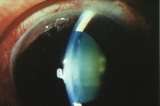
Acute angle closure typically presents with severe ocular pain, headache, blurred vision, halos around lights, nausea, and vomiting. Some of the apparent non-ocular manifestations (nausea/vomiting) could be misleading to the inexperienced physician. However, the prompt recognition and subsequent treatment of an acute angle closure crisis is paramount in the preservation of the patient’s vision. Typical eye exam findings, as in this patient, include mild conjunctival injection, hazy cornea, mid dilated pupil, shallow angle and elevated intraocular pressure.
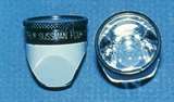
Angle closure is best observed using a gonioscopic contact lens that allows the viewer to see into the angle of the eye. Even if the acute episode is diagnosed and treated quickly and appropriately, there can still be optic nerve damage and resultant visual loss. Another possible change includes iris ischemia which can cause sloughing of iris pigment. Pigment can be noted in the anterior chamber and on the corneal endothelium. Iris damage may cause the pupil to remain in a dilated position. The intraocular pressure may also rise enough to cause retinal vascular occlusion and retinal ischemia. Anterior subcapsular lens opacities may also occur as a result of ischemia (this is termed glaukomflecken).
Treatment:
Treatment of acute angle closure glaucoma is either laser or surgical peripheral iridectomy (placing a hole in the peripheral iris). This procedure restores aqueous flow from the posterior to anterior chamber by creating an extra opening in the iris, relieving the pathologic pressure gradient. This ultimately allows the iris to regress and pull away from the trabecular meshwork and then normal aqueous humor drainage is restored. This procedure is often curative of the affected eye. A prophylactic peripheral iridectomy of the non-affected eye is necessary to prevent an episode from occurring in the fellow eye. Individuals with one episode of acute angle closure glaucoma have a high likelihood of an attack in the fellow eye over the next 5-10 years. Even when the intraocular pressure has decreased, subsequent follow up is necessary to be sure that the angle remains open. IOP may decrease soon after the attack due to ciliary body ischemia and decreased aqueous humor production and not because the angle has reopened.
Medical College of Wisconsin Ophthalmology and Visual Sciences Case Studies
-
Ophthalmic Case Study 1
Acute right eye pain -
Ophthalmic Case Study 2
Red, itchy eyes -
Ophthalmic Case Study 3
Acute left eye pain and blurry vision -
Ophthalmic Case Study 4
Left eye pain and fuzzy vision 2 days after eye surgery -
Ophthalmic Case Study 5
Girl rubbing her R eye after trauma -
Ophthalmic Case Study 6
Red eye and pain on the left -
Ophthalmic Case Study 7
Vision loss L eye -
Ophthalmic Case Study 8
Crossed eyes -
Ophthalmic Case Study 9
White pupils -
Ophthalmic Case Study 10
Blurry vision in the left eye for 2 weeks -
Ophthalmic Case Study 11
Acute pain and burning in L eye -
Ophthalmic Case Study 12
Blurry vision in both eyes and headaches -
Ophthalmic Case Study 13
"Cannot see well" from left eye -
Ophthalmic Case Study 14
Blurry vision in both eyes -
Ophthalmic Case Study 15
Eye irritation and dryness -
Ophthalmic Case Study 16
2 brief episodes of vision loss in the R eye -
Ophthalmic Case Study 17
Routine eye exam -
Ophthalmic Case Study 18
8-year-old boy, difficulty seeing whiteboard -
Ophthalmic Case Study 19
8-year-old girl, wandering eye and double vision. -
Ophthalmic Case Study 20
Red eyelid lesion in an infant -
Ophthalmic Case Study 21
Female patient with optic nerve abnormality -
Ophthalmic Case Study 22
Left eye pain, tearing, redness, and photophobia -
Ophthalmic Case Study 23
1-year-old male presents with intermittent eye misalignment -
Ophthalmic Case Study 24
Left eye pain, redness, and blurry vision -
Ophthalmic Case Study 25
16-year-old non-verbal male with history of retinopathy -
Ophthalmic Case Study 26
69-year-old female presents for decreased visual acuity
Contact Ophthalmology
For patient care inquires, call us at (414) 955-2020 or use MyChart. Email is for research and education inquiries only.
Eye Institute Location
925 N. 87th St.
Milwaukee, WI 53226
Appointments
(414) 955-2020
Continuing Medical Education
Renee Ware
(414) 955-2086
Medical Education Coordinator
Associate Director of Development - Ophthalmology
Sarah Walker
Refer to Us - Consultation requests
Patient Referral Form (PDF)
Fax to (414) 955-0136
Emergent Requests
Within 48 hours call
(414) 955-2020
Research
Vesper Williams
(414) 955-7862
Advanced Ocular Imaging Program
(414) 955-2647
Eye Institute Executive Director (Administrator)
Shannon Dreier