header-logo
Ophthalmology_Hero Image 2

Case Study 9 - CC: White pupils

all
Patient Visit

Patient History
HPI:

The pediatrician was alarmed when he finally got a chance to evaluate the 2 hour-old newborn and was unable to get the usual red reflex in either eye with the direct ophthalmoscope. The reflection in both eyes was grayish white. The mother remembers having flu-like symptoms in her early pregnancy but attributed it to being a part of normal pregnancy.

Past Ocular History:
None

Ocular Medications:
None

Past Medical History:
Birth history: 38 week gestation, normal spontaneous vaginal delivery, no complications

Surgical History:
None

Past Family Ocular History:
No history of eye disease, blindness or congenital cataracts

Social History:
Will live at home with mother and father. Mother denies smoking, alcohol use or other drug use during pregnancy.

Medications:
None

Allergies:
None

ROS:
Otherwise negative

Ocular Exam

Visual Acuity (cc):
OD: Reacts to light
OS: Reacts to light

IOP (tonoapplantation):
OD: Not tested
OS: Not tested

Pupils:
Equal, round and reactive to light, no APD.

Extraocular Movements:
Full OU. No nystagmus.

Confrontational Visual Fields:
Not performed

External:
Normal, both sides

Slit Lamp:

Lids and Lashes Normal OU
Conjunctiva/Sclera Normal OU
Cornea Clear OU
Anterior Chamber Deep and quiet OU
Iris Normal OU
Lens Dense central opacities OU
Anterior Vitreous No view

Dilated Fundus Examination:

OD Unable to perform due to poor view
OS Unable to perform due to poor view

Diagnosis and Discussion

Diagnosis
Leukocoria

Discussion

Differential Diagnosis:
This is a case of leukocoria, or an abnormal white pupillary reflex, likely due to congenital cataracts. Differential diagnosis of leukocoria includes the following: retinoblastoma, congenital cataracts (from infectious [ex. intrauterine rubella infection] or congenital etiologies [ex. galactosemia, Lowe’s syndrome, familial]), retinopathy of prematurity, persistent hyperplastic primary vitreous, Coat’s disease, familial exudative vitreoretinopathy, retinal detachment, coloboma and corneal opacities.

Definition:
Congenital cataracts can form due to intrauterine infections, metabolic disorders, a malignancy, or a genetic defect. Intrauterine infections that can result in congenital cataracts include rubella (German measles, the most common infectious cause), rubeola, cytomegalovirus, herpes simplex, herpes zoster, poliomyelitis, influenza, Epstein-Barr virus, syphilis, and toxoplasmosis. Metabolic disorders that can cause congenital cataracts include galactosemia and diabetes mellitus. Systemic syndromes such as Lowe’s syndrome (oculocerebrorenal syndrome) or Alport syndrome may also be associated with congenital cataracts. The differential of leukocoria, or a white light reflex, must also include retinoblastoma, the most prominent intraocular malignancy in children.

Examination:
A complete medical history including maternal illness or drug use during pregnancy is very important. Family ocular history of congenital blindness, congenital cataracts, strabismus, or amblyopia should also be addressed. A complete eye exam including visual assessment of each eye alone and an attempt to determine the visual significance of the cataract is necessary. B-scan can be helpful to evaluate the posterior eye to rule out posterior abnormalities. A physical examination to determine signs and/or symptoms of systemic intrauterine-acquired infections is essential.

Treatment:
Cataract surgery is the treatment of choice and should be performed between 4-8 weeks of age to minimize the risk of amblyopia and sensory nystagmus. Cataract extraction with primary posterior capsulectomy and anterior vitrectomy is the procedure of choice due to the high rate of capsular opacification. Most patients are left aphakic and are fitted with a contact lens shortly after surgery. Secondary intraocular lens implantation can be done later in life after the eye has matured. After cataract extraction, patients should be assessed and treated for amblyopia. Life-long follow up is important to maximize visual potential.

Self-Assessment Questions
  1. A baby is brought to the pediatrician for her 4 month-old well baby checkup. Which finding would be concerning and merit an ophthalmology referral?
  2. What would happen if a cataract is removed later than 7yrs?
Self-Assessment Answers

A baby is brought to the pediatrician for her 4 month-old well baby checkup. Which finding would be concerning and merit an ophthalmology referral?
b. mom reports that in pictures the R pupil looks bright orange with the camera flash but not the L pupil - a check of the pupillary reflex with the direct ophthalmoscope confirms her findings
An abnormal red reflex on direct ophthalmoscopy needs urgent referral to ophthalmology.

What would happen if a cataract is removed later than 7yrs?
c. the R and L occipital lobes will develop differently
Development of visual pathways would be uneven since the input from the cataractous eye is blurrier than the other eye.

Contact Ophthalmology

For patient care inquires, call us at (414) 955-2020 or use MyChart. Email is for research and education inquiries only.

Eye Institute Location

925 N. 87th St.

Milwaukee, WI 53226


Appointments

(414) 955-2020

(414) 955-6166 (fax)


Continuing Medical Education

Amanda Tan

atan@mcw.edu

(414) 955-2049


Medical Education Coordinator

Ophth-Residency@mcw.edu


Associate Director of Development - Ophthalmology

Sarah Walker

sarawalker@mcw.edu

Refer to Us - Consultation requests

Patient Referral Form (PDF)

Fax to (414) 955-0136


Emergent Requests

Within 48 hours call

(414) 955-2020


Research

Vesper Williams

vewilliams@mcw.edu

(414) 955-7862


Advanced Ocular Imaging Program

aoip@mcw.edu

(414) 955-2647


Eye Institute Executive Director (Administrator)

Shannon Dreier
sdreier@mcw.edu

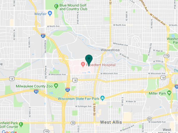
Eye Institute Google map location