
header-logo
Ophthalmology_Hero Image 2

Case Study 28: Recurring bilateral “significant eyelid lesions” following ptosis repair 

Original Authors: Leah Poulos, MD, Smith Ann Meile Chisholm, MD 

all
Patient Visit

HPI
9yo boy presents for evaluation of recurring b/l “significant eyelid lesions.” He was initially seen by a colleague at the age of 2 for b/l upper eyelid drooping present since birth. He returned at the age of 5 for surgical consultation; frontalis sling placement for ptosis repair was recommended but parents wanted to wait and patient was lost to follow up. At the age of 9, he underwent ptosis repair surgery with polytetrafluoroethylene suture slings at an OSH. His post-operative course was c/b development of recurrent b/l upper eyelid lesions refractory to excision in addition to courses of oral prednisone and topical maxitrol.

Past Ocular History
Severe congenital ptosis with poor levator function OU, lateral canthal dystopia OU, intermittent exotropia with BIOOA, myopia with astigmatism OU

Ocular Medications
None

Past Medical History
ASD with accompanying intellectual impairment requiring substantial support (level 2), ADHD (combined type), speech delay, birth history non-contributory (born at 39 weeks via uncomplicated c-section)

Surgical History
Full dental rehabilitation (02/20217), Bilateral ptosis repair with polytetrafluoroethylene suture slings (5/2022)

Past Family Ocular History
Glaucoma and refractive error in mother, refractive error in father

Social History
Lives with mom and dad. No smoking in the house.

Medications
Cetirizine 10 mg daily

Allergies
NKDA

ROS
Patient’s mother reports “bumps get bigger all the time” and will itch and bleed, otherwise negative


Ocular Exam

Visual Acuity (cc)
OD:20/30 -2
OS:20/40 -2

IOP (iCare tonometry)
OD: 14 mmHg
OS: 15 mmHg

Pupils
OD: 5 > 3, brisk round, reaction to light, no APD
OS: 5 > 3, round, brisk reaction to light, no APD

Extraocular Movements
OD: see sensorimotor exam for details
OS: see sensorimotor exam for details

Confrontational Visual Fields
OD: Full
OS: Full

Slit Lamp:

OD OS
External Incisions well healed with small lesion at medial brow incision Incisions well healed with no lesions present
 MRD1 -2 mm 1 mm
Palpebral Fissure 5 mm 5 mm
Lids and Lashes 3x2 mm nodule UL nasal along eyelid crease incision, ptosis 4x6 mm nodule UL nasal along eyelid crease incision, ptosis
Conjunctiva/Sclera White and quiet White and quiet
Cornea Clear Clear
Anterior Chamber Deep and quiet Deep and quiet
Iris Normal Normal
Lens Clear Clear
Anterior Vitreous Normal Normal

Dilated Fundus Examination:

OD OS
Disc Normal Normal
Macula Normal Normal
Vessels Normal Normal

Imaging/Additional Tests

Sensorimotor exam:
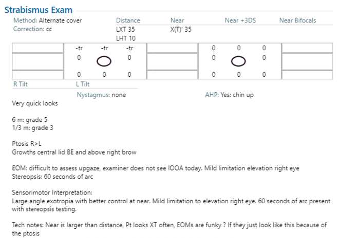
Sensorimotor Exam


Diagnosis and Discussion

Diagnosis
Pyogenic granuloma

Differential Diagnosis
The most likely diagnosis for this patient is bilateral pyogenic granulomas in the setting of prior bilateral ptosis repair surgery. During his bilateral upper eyelid excision, right brow lesion excision and removal of bilateral polytetrafluoroethylene slings, it was noted that the slings were in direct communication with the lesions. In addition, the surgical pathology showed granulation tissue that likely represented an inflammatory reaction to the foreign body. Alternative diagnoses that should be considered and ruled out include chalazion, hordeorum, angioendothelioma, angiosarcoma, neurofibroma, hemangioendothelioma, squamous cell carcinoma and intravascular papillary endothelial hyperplasia.

Definition
A pyogenic granuloma is a rapidly growing benign vascular lesion involving the skin or mucosa (e.g., cornea, conjunctiva, eyelids) composed of granulation tissue and non-granulomatous inflammatory cells. Their name is misleading, as they are neither pyogenic (pus-producing) or granulomatous. They are often seen at the site of previous trauma, surgery or inflammation and will bleed spontaneously or from light touch. Their etiology is unclear but thought to be affected by angiogenic dysregulation- more specifically, disordered fibroblast proliferation and formation of capillaries radiating in lobules, which is why they are also known as lobular capillary hemangioma. Proliferation of capillaries and granulation tissue during wound healing is usually transient, lasting a few weeks until epithelial tissue has been fully repaired; whereas, in the case of pyogenic granulomas, it appears that excessive endothelial proliferation occurs due to ongoing tissue irritation and/or inflammation. In the setting of tissue injury, it has been suggested there may be activation of a neovascular pathway involving FLT4 and nitric oxide in addition to overexpression of VEGF, mutations in BRAF and RAS and development of tumorigenic transcription factors including STAT3. They are a known potential complication of multiple oculoplastic surgical procedures such as chalazion curettage, blepharoplasty, and ptosis repair (especially Frontalis Sling Placement using non-autogenous material), with an incidence ranging from 9-25%. They predominately arise 1-6 weeks after surgery, with most cases occurring within the first 2-4 weeks post-operatively (as this timeline is consistent with the proliferative phase of wound healing). In a study reviewing 100 cases of ocular pyogenic granuloma, approximately 42% were due to chalazion, 40% were following ocular surgery (e.g., pterygium excision, strabismus surgery, nasolacrimal duct probing, enucleation) and 5% were related to accidental trauma.

Examination/diagnostics
Preliminary diagnosis is clinical and made in the setting of a rapidly growing (weeks to months), smooth, painless but friable papule or nodule that may show purulence and crusting. They may cause some discomfort or irritation depending on location and size. On patient presentation, the average size is around 6.5 mm and maximum size is 1 cm. Grossly, pyogenic granulomas are fleshy pink/red/purple hemorrhagic masses that can be flat, polypoid or pedunculated seen on the lids/conjunctiva/cornea, often near an incision site in post-operative cases. Definitive diagnosis is made through histopathologic examination using hematoxylin and eosin (H&E) stain following biopsy of the lesion and is essential to rule out conditions that mimic pyogenic granuloma, especially malignancies. On histology, pyogenic granulomas appear with branching endothelium-lined vessels and prominent lobular capillary proliferation, dense non-granulomatous inflammatory cell infiltration, granulation tissue and/or increased fibromyxoid stromal fibrosis. The overlying epidermis may show ulceration, hyperkeratosis, or hypergranulosis. Due to its vascular origin, immunohistochemical stains including CD34, CD31, Factor VIII, and SMA are positive.

Treatment
If untreated, some spontaneously regress and atrophy. Conservative management, which has been observed to be largely effective in smaller lesions, includes topic corticosteroids and beta blockers as first-line pharmacotherapy. For cases involving the conjunctiva, management with steroid or bleomycin/bevacizumab injections, sclerosis agents (e.g., polidocanol), Imiquimod or non-surgical cryotherapy with liquid nitrogen may be used. Larger lesions and/or refractory cases often require surgical intervention (excision of lesion at its base, curettage, electrothermal cauterization, shave excision with laser, blunt removal with chemicals). Given the possibility of malignancy (prior to definitive diagnosis), complete surgical excision is preferred, as it yields a specimen for pathologic examination. Consideration of management should also include optimal cosmetic outcomes depending on lesion location. Overall, recurrence rates are lowest with surgical excision and cryotherapy, estimated to be around 2.94% and 1.62%, respectively. Recurrence is more common when there is extension of vasculature within the dermis.


Self-Assessment Questions
  1. Which is not a treatment modality for pyogenic granuloma?
    a. Complete surgical excision
    b. Topical 5-fluorouracil (5-FU)
    c. Intralesional bleomycin
    d. Topical timolol
    e. Topical prednisolone

  2. A 4-year-old girl is brought to clinic by her father for her two week follow up examination for unilateral medial rectus recession and lateral rectus resection. Her post-operative course has been uncomplicated except for a painless 3 mm red-purple bump that has been growing in size for the past 4 days near her incision site. After taking a thorough history and performing an exam, what is the next best step in management?
    a. Do nothing, the bump is normal post-operatively and will go away on its own.
    b. Have the family come back in one week if the bump is still there for further evaluation.
    c. Schedule surgical excision immediately, as there is high concern for malignancy.
    d. Prescribe topical 0.5% timolol twice daily for six weeks and have them return to clinic for re-evaluation.
Self-Assessment Answers
  1. Which is not a treatment modality for pyogenic granuloma?
    b. Topical 5-fluorouracil (5-FU)

    5-fluorouracil (5-FU) is not a treatment modality for pyogenic granuloma. It is used in the treatment of squamous cell carcinoma, which is an important differential diagnosis as both conditions can present with rapidly growing, fleshy friable nodules on the skin/mucous membranes. Interestingly, 5-FU has been associated with the development of pyogenic granuloma.

    Curr N., Saunders H., Murugasu A., Cooray P., Schwarz M., and Gin D. (2006). Multiple periungual pyogenic granulomas following systemic 5-fluorouracil. Australas J Dermatol. May;47(2):130-3. doi: 10.1111/j.1440-0960.2006.00248.x. PMID: 16637811.

    McDonald S. M. and Neidenbach P.J. (2024). Intralesional 5-Fluorouracil in the Treatment of Squamous Cell Carcinoma in an Elderly Patient. Cureus. 2024;16(3):e55855. DOI:10.7759/cureus.55855


  2. A 4-year-old girl is brought to clinic by her father for her two week follow up examination for unilateral medial rectus recession and lateral rectus resection. Her post-operative course has been uncomplicated except for a painless 3 mm red-purple bump that has been growing in size for the past 4 days near her incision site. After taking a thorough history and performing an exam, what is the next best step in management?
    d. Prescribe topical 0.5% timolol twice daily for six weeks and have them return to clinic for re-evaluation.


    Prescribe topical 0.5% timolol twice daily for six weeks and have them return to clinic for re-evaluation. This is the best next step in management for this patient who is 2 weeks post-op from strab surgery, which is a common predisposing factor to pyogenic granuloma formation. Because her bump is only 3 mm, the best option is a trial of conservative management with topical timolol (could also choose topical steroids or a combination). As clinical suspicion for the diagnosis of pyogenic granuloma is high, it is not appropriate to do nothing (A, B). However, conservative treatment should be trialed first (D) before more invasive options (C) are considered.

    Barsegian, A., Silverman, N., and Shinder, R. (2018). Ocular Pyogenic Granuloma Treated with Topical Timolol. Investigative Ophthalomology & Vision Science, July; 59(9). https://iovs.arvojournals.org/article.aspx?articleid=2694662


References/Resources

  1.  Belliveau, M. J., and DeAngelis, D. D. (2016). Nonhealing traumatic wound over frontalis silicone sling. Canadian Journal of Ophthalmology, 51(3), E88-89. DOI: 10.1016/jcjo.2016.01.008.
  2. Cheung, I., and Cheung, D. (2026). Postoperative Eyelid Pyogenic Granuloma: A Systematic Review of Clinical Features, Surgical Associations, and Management. Cureus, 18(5):e108239. DOI: 10.7759/cureus.108239.
  3. Dlaim, M. S. B., Alhussein, G. A., Alqahtani, R. S., and Almanea, L. T. (2023). Conservative Management of Giant Pyogenic Granuloma Post Strabismus Surgery: Case Report and Literature Review. Cureus, 15(7):e41321. DOI: 10.7759/cureus.41321.
  4. Kaleeny, J. D., and Janis, J. E. (2024). Pyogenic Granuloma Diagnosis and Management: A Practical Review. Plastic and Reconstructive Surgery Global Open, 12(9):e6160. DOI: 10/1097/ GOX.0000000000006160
  5. Mehta, A., Naik, M., Abrol, S., Garg, P., and Joshi, M. (2017). Granuloma after sling surgery: an attempt to answer the ‘why’ and ‘what to do next.’ International Ophthalmology, 37, 973-977. DOI: 10.1007/s10792-016-0342-0.
  6. Phelps, P., Wells, J., Yen, M. T., Nguyen, B. J., and Thyparampil, P. (last updated August 2026). Pyogenic Granuloma. American Academy of Ophthalmology: EyeWiki. https://eyewiki.org/Pyogenic_Granuloma.
  7. Ramirez, D. A., Stiff, H. A., and Greiner, M. A. Pyogenic Granuloma. EyeRounds.org. Available from: https://webeye.ophth.uiowa.edu/eyeforum/atlas/pages/pyogenic-granuloma/index.htm#gsc.tab=0.
  8. Yoshida, H., Tominaga, S., and Makino, S. (2020). Conjunctival Pyogenic Granuloma. The Journal of Pediatrics: X, 4:100035. DOI: 10.1016/j.ympdx.2020.100035.