header-logo
Ophthalmology_Hero Image 2

Case Study 27: Lesion on the surface of the left eye

Original Authors: Rohit Balaji, Heather Stiff, MD

all
Patient Visit

HPI
A 9-year-old female presented to the ophthalmology clinic for follow up of a lesion on the ocular surface of left eye (OS) which has been present since birth. She has been following with pediatric ophthalmology for this lesion and left exotropia. She notes poor vision in the left eye but has had no recent vision changes. She wears glasses full time for protection.

Past Ocular History

  • Ocular Surgery
    • Underwent multiple excisions of left ocular surface lesion with amniotic membrane grafts
    • Removal of left eyelid lesions
  • Ocular Diagnoses
    • Mechanical lagophthalmos of OS
    • Retinal/choroidal hamartoma of OS
    • Optic nerve hypoplasia OS
    • Anisometropic and deprivation amblyopia OS
    • Sensory exotropia of OS

Ocular Medications
Nightly lubricating ointment in OS

Past Medical History
Significant for Haberland syndrome, epilepsy, right hemiparetic cerebral palsy, and aplasia cutis

Surgical History
History of left hemispherectomy

Past Family Ocular History
None

Social History
Doing well in elementary school

Medications
Clobazam 10mg daily
Diazepam 10mg as needed (PRN) for seizures
Lacosamide 150mg daily
Tacrolimus ointment PRN skin dryness
Botox injections for spasticity

Allergies
No known allergies (NKA)

ROS
As per HPI, otherwise negative

Ocular Exam

Visual Acuity (cc)
OD:20/50 -1
OS:20/600

IOP (iCare tonometry)
OD: 20 mmHg
OS: 17 mmHg

Pupils
OD: 6mm in dark, 4mm in light, Round, Brisk no APD
OS: 6mm in dark, 4mm in light, Round, Brisk no APD

Slit Lamp:

OD OS
External Normal Dryness/scaling of periorbital skin, soft nodule at left lateral canthus 
Lids and Lashes Normal Lagophthalmos < />
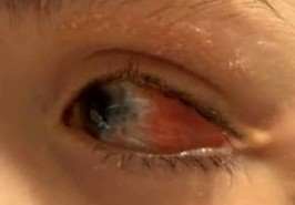
Conjunctiva/Sclera White and quiet Residual yellow-white lesion temporally with superotemporal scleral thinning near limbus, chemosis and injection over scar temporally  
Cornea Clear Central and inferior scarring/pannus, conjunctivalization temporally and nasally
Anterior Chamber Deep and quiet Deep and quiet
Iris Round and Reactive Round and Reactive
Lens Clear Clear
Anterior Vitreous Normal Normal

Dilated Fundus Examination:

OD OS
Disc Normal Small, hypoplastic
Macula Normal Normal, foveal reflex is present
Vessels Normal Normal
Periphery Normal Choroidal nevus vs hamartoma

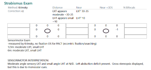
Strabismus Exam

Retinoscopy: OD: +0.50 +1.75 X 095, OS: +4.00 +4.50 X 120
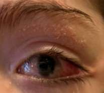
Retinoscopy ODRetinoscopy OS

Figure 1. Appearance of ocular surface and periorbital region


Diagnosis and Discussion

Diagnosis
Epibulbar/limbal dermoid of the left eye in setting of Haberland syndrome

Discussion

  • Differential Diagnosis
    • Pterygium
      • A pterygium is a growth of wing-shaped fibrovascular tissue at the limbus extending onto the corneal surface, usually nasally.1 Pterygia are generally driven by cumulative UV exposure and, thus, would be very atypical in a 9-year-old. Additionally, pterygia appear as pink, vascular structures rather than opaque and yellow-white as an epibulbar dermoid, further steering away from a pterygium as a diagnosis.
    • Pinguecula
      • Like pterygia, a pinguecula is a benign, acquired lesion associated with cumulative UV exposure. Pingueculae are confined to the bulbar conjunctiva and do not cross the limbus onto the cornea like pterygia.2 This is likely not the right diagnosis in this case for similar reasons as a pterygium.
    • Conjunctival nevus
      • Conjunctival nevi are melanocytic tumors that are typically small, isolated lesions near the limbus.3 They can be either pigmented or non-pigmented (amelanotic), often with intrinsic cysts. The lesion in this case, however, was a larger solid limbal mass in the presence of other eye abnormalities, which argues against a diagnosis of a conjunctival nevus.
    • Conjunctival inclusion cyst
      • Conjunctival inclusion cysts are benign cysts that are filled with clear serous fluid or mucoid material. While it could be considered in the differential diagnosis, it is quickly ruled out as the lesion in this case was solid, opaque, and not fluid filled.
    • Conjunctival papilloma
      • Conjunctival papillomas are acquired epithelial tumors and have a classic finger-like or cauliflower-like appearance.4 They are often lobulated and have a central vascular core. A diagnosis of a conjunctival papilloma is less likely in this case as the lesion did not have a cauliflower-like appearance and was congenital in nature, rather than acquired.

Definition
In general, dermoid lesions are benign congenital choristomas that arise from ectodermal and mesodermal tissue that gets trapped along embryonal fusion lines during fetal development.5 On histopathology, they are usually lined by keratinized epithelium and can contain tissue such as hair follicles, sebaceous glands, sweat glands, or cartilage.5

Dermoids can be separated into three main subtypes based on their clinical appearance and location. The first type is an epibulbar/limbal dermoid, which is the diagnosis for the patient in this case. Limbal dermoids typically have a firm whitish-yellow dome-like appearance classically at the inferotemporal corneal limbus. On examination, they may have fine hair growing from the surface. The second type is a lipodermoid. These are characterized as soft, yellowish-pink fatty masses usually found in the superotemporal fornix. The third type is an orbital dermoid. These are among the most common orbital tumors of childhood and typically present as a painless, subcutaneous cystic mass at the orbital rim.

Epibulbar/limbal dermoids can cause substantial astigmatism from corneal distortion and can lead to significant anisometropia. Large limbal dermoids can also encroach onto the visual axis. Both anisometropia and visual axis obscuration can lead to amblyopia of the affected eye. This, in turn, can lead to further complications such as sensory strabismus. These findings were seen in the affected eye of the patient in this case, though some of the amblyopia in this case is also attributed to a hypoplastic optic nerve. Additional complications can include ocular surface irritation from hairs present on the dermoids, cosmetic concerns, and rupture of cysts in orbital dermoids.

Dermoid lesions can occur in isolation or in association with systemic syndromic conditions. Such conditions include Goldenhar syndrome, linear nevus sebaceous syndrome, or encephalocraniocutaneous lipomatosis/Haberland syndrome, the last of which the patient in this case was diagnosed with. Briefly, Haberland syndrome is a rare neurocutaneous disorder presenting with ocular, dermatologic, and CNS lesions.6,7 It is characterized by a hallmark lesion known as nevus psiloliparus and craniofacial lipomas. Ocular findings can include epibulbar dermoids, lipodermoids, corneal or scleral defects, anterior chamber irregularities, aniridia, microphthalmia, and periocular skin tags.6,7 Neurological features of Haberland syndrome may include seizures, developmental delays, and CNS lipomas.6,7

Examination/diagnostics
Diagnosis is largely clinical and warrants a thorough history and physical examination including visual acuity, sensorimotor examination, and cycloplegic refraction. Diagnostic imaging, including anterior segment ultrasound biomicroscopy or anterior segment ocular coherence tomography (OCT), can be used to determine depth of limbal dermoids.

Treatment
Treatment of epibulbar dermoids depends on the grade of the lesion, which is determined by the depth and extent of the lesion.8 Grade I lesions involve the superficial cornea and may be managed with observation if small, stable, and not visually significant, or with simple excision. Deeper Grade I lesions may warrant excision and grafting with amniotic membrane or limbal stem cell transplant to decrease the risk of pseudopterygium development or corneal neovascularization.9

Grade II lesions involve the full thickness of the cornea with or without corneal endothelial involvement. Treatment for these lesions often requires a lamellar keratoplasty.9

Grade III lesions involve the entire cornea and invade into the anterior chamber. These lesions typically require penetrating keratoplasty and complex anterior segment reconstruction.9

Complications following surgery may include corneal perforation, corneal scarring, pseudopterygium, corneal conjunctivalization from limbal stem cell deficiency, and graft rejection.9 Additionally, patients may require multiple staged procedures for complete treatment. Despite treatment, astigmatism and amblyopia may persist, requiring long-term vision therapy.


Self-Assessment Questions
  1. What clinical feature is characteristic of a limbal dermoid?
    a. Soft, fatty mass in the superotemporal fornix
    b. A cystic mass filled with clear serous or mucoid fluid
    c. Firm, whitish-yellow lesion at the inferotermporal corneal limbus
    d. Pigmented lesion with cysts arising at corneal limbus

  2. What is the correct treatment approach for a Grade II limbal dermoid?
    a. Lamellar keratoplasty
    b. Superficial excision with amniotic membrane graft
    c. Enucleation of the affected eye
    d. Laser ablation of the lesion

Self-Assessment Answers
  1. What clinical feature is characteristic of a limbal dermoid?

    c. Firm, whitish-yellow lesion at the inferotermporal corneal limbus

    Limbal dermoids typically appear as firm, whitish-yellow dome shaped lesions usually at the inferotemporal corneal limbus. They may contain fine hair on examination.

  2. What is the correct treatment approach for a Grade II limbal dermoid?
    a. Lamellar keratoplasty

    Grade II limbal dermoids involve the full thickness of the cornea, with or without corneal endothelial involvement and often require lamellar keratoplasty for treatment.


References/Resources:

  1. Hirst L, Bunya VY, Wood MA, et al. Pterygium. EyeWiki. Accessed August 27, 2026. https://eyewiki.org/Pterygium.
  2. Al-Hashimi S, Bunya VY, Woodward MA, Halfpenny C. Pinguecula. EyeWiki. August 11, 2026. Accessed August 27, 2026. https://eyewiki.org/Pinguecula.
  3. Yeager LB, Kassotis A, Frank T, Li CY, Marr BP. A review of pediatric ophthalmic tumors. Pediatrics in Review. 2024;45(3):119-131. doi:10.1542/pir.2023-006163
  4. Phelps P, Duong H-VQ, Goel S, et al. Conjunctival Papilloma. EyeWiki. Accessed August 27, 2026. https://eyewiki.org/Conjunctival_Papilloma.
  5. Mansour AM, Barber JCK, Reinecke RD, Wang F. Ocular Choristomas. Survey of Ophthalmology. 1989;33(5):339-358.doi:https://doi.org/10.1016/0039-6257(89)90011-8
  6. Moog U, Dobyns WB. Encephalocraniocutaneous Lipomatosis. 2022 Jan 27. In: Adam MP, Bick S, Mirzaa GM, et al., editors. GeneReviews® [Internet]. Seattle (WA): University of Washington, Seattle; 1993-2026. Available from: https://www.ncbi.nlm.nih.gov/books/NBK576966/
  7. Lopes, F. C. P. S., Schroeder, C., Patel, B., & Levy, M. L. (2024). Review of encephalocraniocutaneous lipomatosis. Seminars in Pediatric Neurology, 52, 101166. https://doi.org/10.1016/j.spen.2024.101166
  8. Pirouzian A. Management of pediatric corneal limbal dermoids. Clinical Ophthalmology. 2013;7:607.doi:https://doi.org/10.2147/opth.s38663
  9. Lang SJ, Böhringer D, Reinhard T. Surgical management of corneal limbal dermoids: retrospective study of different technique sand use of Mitomycin C. Eye (Lond). 2014;28(7):857-862. doi:10.1038/eye.2014.112